Detection and analysis of spatially varying fluid levels using magnetic signals

a magnetic signal and fluid level technology, applied in the field of noninvasive, diagnostic, medical devices, systems and methods, can solve the problems of not having continuous monitoring technology available to alert clinical staff, many brain injuries not severe enough to warrant drilling a hole in the cranium, and no continuous monitoring technology

Active Publication Date: 2015-12-31
CEREBROTECH MEDICAL SYST
View PDF9 Cites 41 Cited by
  • Summary
  • Abstract
  • Description
  • Claims
  • Application Information

AI Technical Summary

Benefits of technology

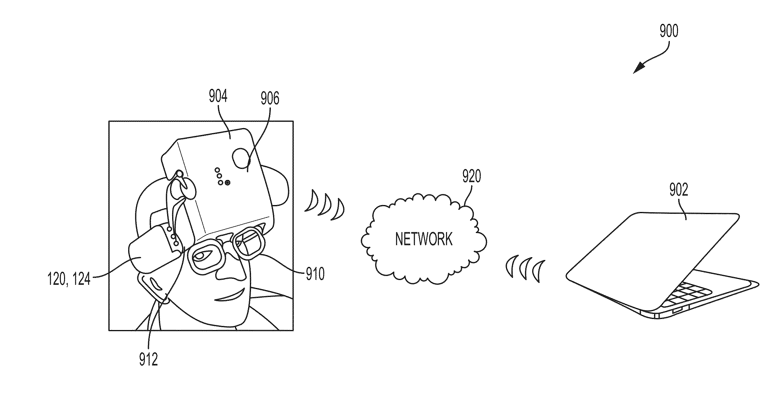
[0010]In yet another embodiment, the present disclosure includes a method for detecting variations in fluid levels in a patient. The method includes attaching a headset to the patient, the headset including a support band for securing the headset to a head of the patient, a processing element coupled to the support band and configured to transfer data wirelessly to an external computer, and multiple transmitter receiver components operably connected to the support band at discrete locations. The method further includes activating the headset to take one or more fluid level readings within the head of the patient, wirelessly transmitting the fluid data corresponding to the one or more fluid level readings from the processing element to the external computer, and analyzing the fluid data with the external computer.

Problems solved by technology

No continuous, noninvasive measurement techniques are currently commercially available for detecting cerebral fluid changes, such as those that occur with bleeding or edema.
Furthermore, many brain injuries are not severe enough to warrant drilling a hole in the cranium for invasive monitoring.
Thus, for many patients with brain injury, there is no continuous monitoring technology available to alert clinical staff when there is a potentially harmful increase in edema or bleeding.
In other words, there is no way currently available for monitoring intracranial fluid changes themselves, and thus the ability to compensate for such changes is limited.
However, no practical, mass-produced medical device based on VIPS technology has yet emerged, to provide clinicians specializing in brain treatment or other areas of medicine the promised benefits of such a device.

Method used


Image

  • Detection and analysis of spatially varying fluid levels using magnetic signals
  • Detection and analysis of spatially varying fluid levels using magnetic signals
  • Detection and analysis of spatially varying fluid levels using magnetic signals

Examples


experimental example

[0171]This experiment was based on the idea that substantial insight can be found in the electromagnetic signature response to a voluntary change in tissue condition. This could lead to a much more controlled diagnostic method, based on electromagnetic measurements of biological tissue condition. In our experiment, a voluntary change was produced in the interrogated organ or tissue, and the diagnostics were performed by evaluating the changes of electromagnetic properties that occurred in those organs or tissue in response to the voluntary produced change and correlating these changes to the voluntary action.

[0172]One example of the method relates to brain concussion, an important medical problem in sports medicine. Sports induced concussion or mild traumatic brain injury (mTBI) is of increasing concern in sports medicine. Neuropsychological examination is the main diagnostic tool for detecting mTBI. However, mTBI also produces physiological effects that include changes in heart rat...


PUM


Abstract

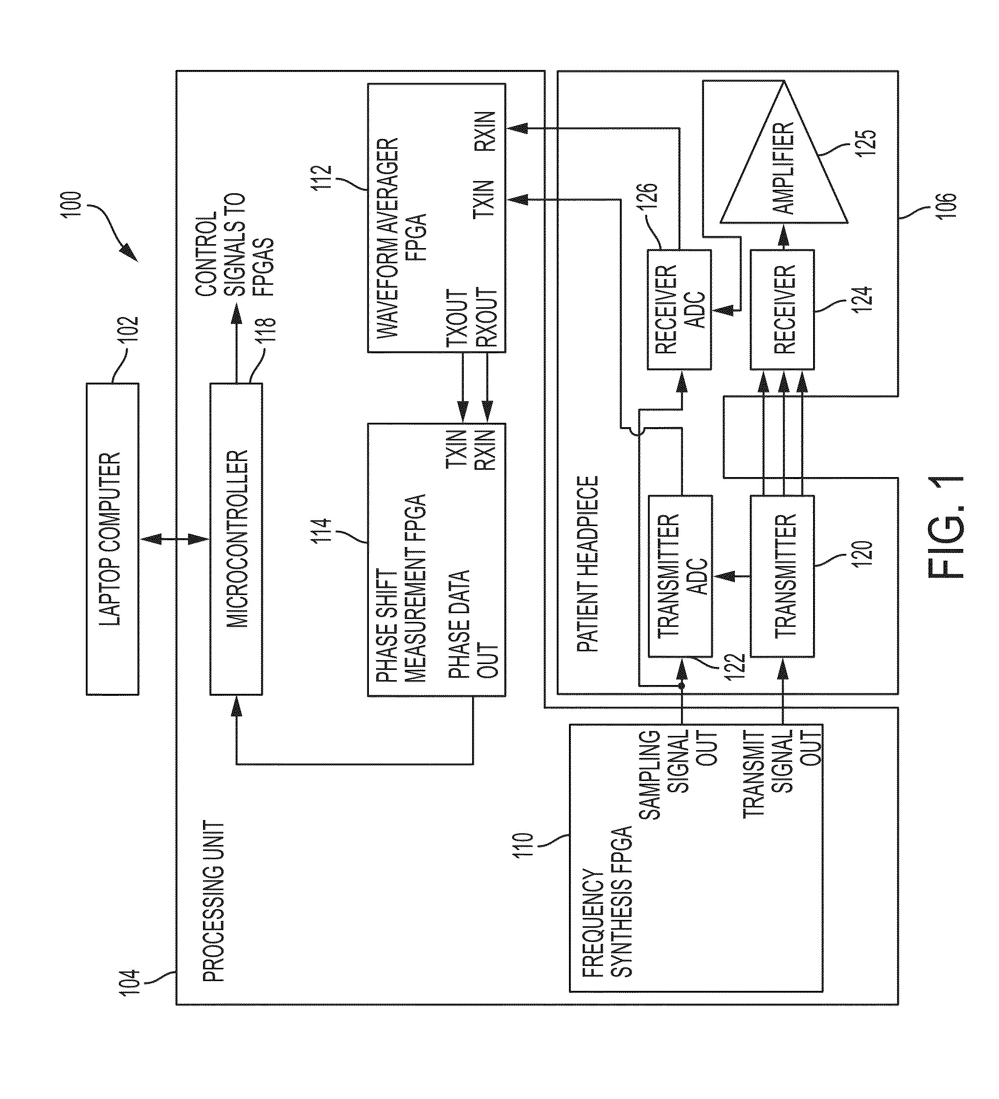
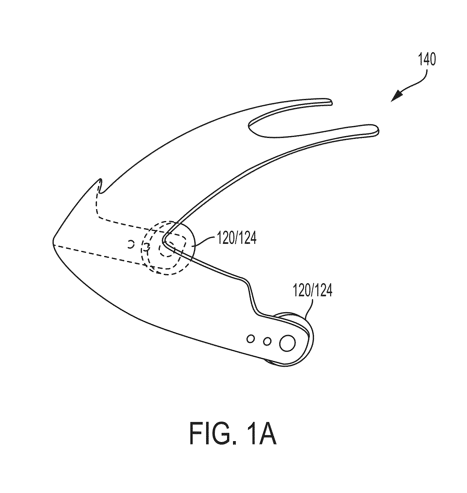
A device for detecting spatial differences in fluid level changes in a tissue of a patient may include a support structure for securing the device to a body part of the patient, a processing element operably connected to the support structure, a wireless networking interface operably connected to the support structure and in communication with the processing element and an external computing device via a network, a first transmission module operably connected to the support structure and in communication with the processing element, a second transmission module and a third transmission module operably connected to the support structure and in communication with the processing element. When activated, the first transmission module transmits a first time varying magnetic field through the tissue of the patient. The second and third transmission modules, which are spatially separated from one another, receive first and second versions, respectively, of the first time varying magnetic field.

Description

CROSS-REFERENCE TO RELATED APPLICATIONS[0001]This application is a continuation-in-part of U.S. non-provisional patent application Ser. No. 14 / 690,985, entitled “Method for Detecting and Treating Variations in Fluid,” filed Apr. 20, 2015, which is a continuation of U.S. non-provisional patent application Ser. No. 14 / 275,549, entitled “Method for Detecting and Treating Variations of Fluid,” filed May 12, 2014, which is a continuation of U.S. non-provisional patent application Ser. No. 13 / 745,710, entitled “Diagnostic method for detection of fluid changes using shielded transmission lines as transmitters or receivers,” filed Jan. 18, 2013, and issued as U.S. Pat. No. 8,731,636 on May 20, 2014, which claims benefit of U.S. provisional patent application Ser. No. 61 / 588,516, entitled “Diagnostic Device for Detection of Fluid Changes in the Brain and Other Areas of the Body,” filed Jan. 19, 2012.[0002]This application also claims priority to U.S. provisional patent application Ser. No. 6...

Claims


Application Information

Patent Timeline
Patent Type & Authority Applications(United States)
IPC IPC(8): A61B5/00A61B5/0452A61B5/03A61B5/04
CPCA61B5/721G16H50/30A61B5/0042A61B5/05A61B5/4064A61B5/4839A61B5/4848A61B5/4878A61B5/6814A61B5/7225A61B5/7275A61B2562/166A61B2562/182A61B2562/222A61B2576/026A61B5/7257A61B5/4875A61B5/0022A61B5/6803A61B5/031A61B2562/0223A61B2560/0223A61B5/04008A61B5/0013A61B5/7214A61B5/0522A61B6/032A61B5/7282G16H40/67G16H50/20A61B5/245
Inventor WYETH, RICHARD WARRENLEVINSON, MITCHELL ELLIOTTWEBER, BRYAN JONSHUSTERMAN, EUGENE MARKRUBINSKY, BORIS
Owner CEREBROTECH MEDICAL SYST
Why Eureka
Try Eureka