VISTA Antagonist and Methods of Use

a technology of vista and antagonist, applied in the field ofvista antagonist and methods of use, can solve the problems of systemic autoimmune diseases, inability of ctla-4 ko mice to adequately control inflammation,

Inactive Publication Date: 2016-03-24
NOELLE RANDOLPH J +4
View PDF6 Cites 30 Cited by
  • Summary
  • Abstract
  • Description
  • Claims
  • Application Information

AI Technical Summary

Benefits of technology

[0021]In one embodiment, the isolated VISTA or PD-1 or PD-L1 or PD-L2 agonist combination elicits a synergistic or additive effect on autoimmune, allergic or inflammatory activity.

Problems solved by technology

(1993) Science 262, 907-909), whereas CTLA-4 KO mice can not adequately control inflammation and develop systemic autoimmune diseases.

Method used


Image

  • VISTA  Antagonist and Methods of Use
  • VISTA  Antagonist and Methods of Use
  • VISTA  Antagonist and Methods of Use

Examples


example 1

Enhancement of T Cell Proliferation

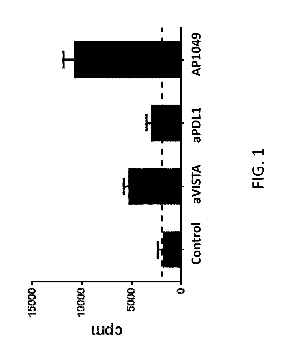
[0177]VISTA+CD11b+ monocytes were enriched from naïve splenocytes using CD11b magnetic beads (Miltenyi). VISTA+CD11bhi MHCII+ myeloid APCs were FACS sorted, irradiated (2500 rads), and used as antigen-presenting cells to stimulate OT-II transgenic CD4+ T cells in the presence of OVA peptide. Control-Ig, monoclonal antibody specific for VISTA and PD-L1 (30 μg / mL), or VISTA-specific peptide (100 μg / mL) were added as indicated. Cell proliferation was measured by tritium incorporation during the last 8 hours of a 72-hour assay. This analysis indicated that T cell proliferation was enhanced in the presence of VISTA or PD-L1 neutralizing monoclonal antibodies, or the AP1049 peptide (FIG. 1). In fact, the AP1049 peptide stimulated T cell proliferation much better than either of the monoclonal antibodies, indicating that the peptide possesses strong antagonistic activity against VISTA.

example 2

Enhancement of T Cell Proliferation

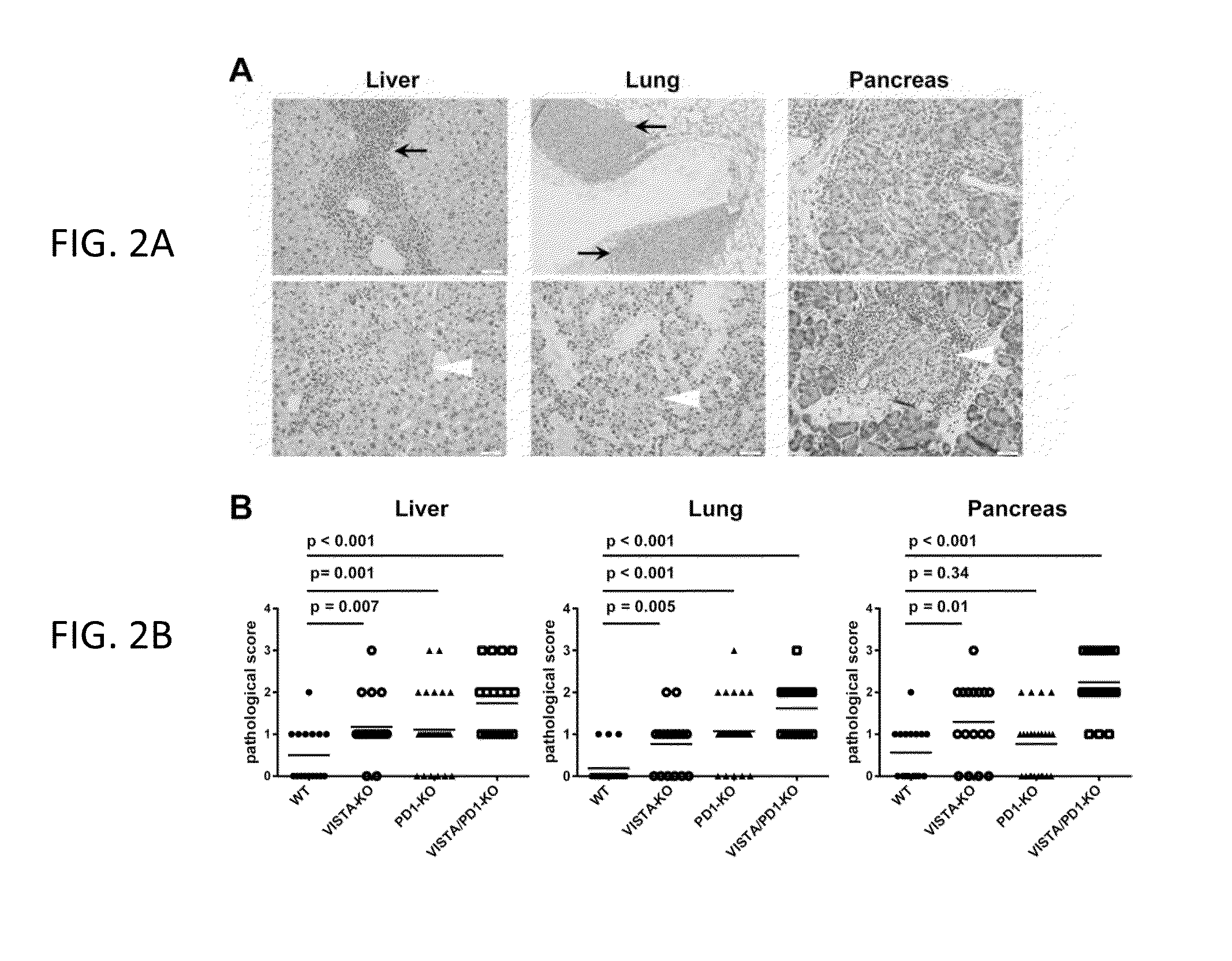
[0178]This example relates to the experiment in FIG. 2. Histological analysis of aged VISTA KO, PD-1 KO, and VISTA / PD-1 double KO mice. Necropsy was performed on 12 months old WT (n=16), VISTA KO (n=15), PD-1 KO (n=28), and VISTA / PD-1 double KO (n=25) mice. Organs were fixed, paraffin embedded, sectioned, and stained with H&E. Two representative H&E sections from lung, liver, and pancreas of the VISTA / PD-1 double KO mice were shown in (A). Clusters of tissue-infiltrating leukocytes were marked with black arrows. (Top row) Areas of necrotic tissues were marked with white arrows (Bottom row). All images are of 200× magnification. Scale bar: 50 microns. The inflammatory state of the tissues was evaluated based on a semi-quantitative method that scores the level of the leukocyte infiltration and tissue necrosis (B).

example 3

Spontaneous T Cell Activation in the VISTA KO, PD-1 KO, and VISTA / PD-1 Double KO Mice

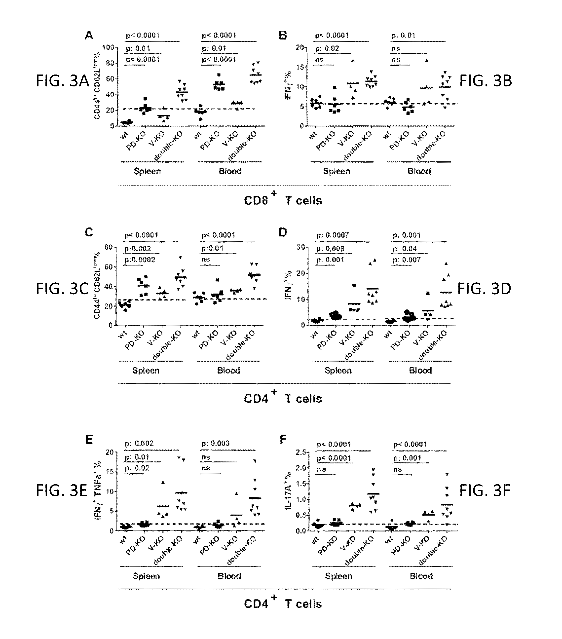
[0179]This example relates to the experiments in FIG. 3 showing spontaneous T cell activation in the VISTA KO, PD-1 KO, and VISTA / PD-1 double KO mice. Splenic T cells were collected from age and gender-matched 6-7 months old WT (n=6), VISTA KO (n=4), PD-1 KO (n=6), and VISTA / PD-1 double KO (n=8) mice. The percentages of CD8+ and CD4+ T cells with activated phenotype (CD44hi CD62Llo) were quantified by flow cytometry. T cells were stimulated ex vivo overnight with soluble anti-CD3 / CD28 mAbs, and their cytokine production (i.e. IFNγ, TNFαβγ and IL-17A) was examined by intracellular staining. CD8+ T cell phenotypes were shown in A and B. CD4+ T cell phenotypes were shown in C-F. Representative results of at least three independent experiments were shown.


PUM

PropertyMeasurementUnit
pHaaaaaaaaaa
compositionaaaaaaaaaa
NMRaaaaaaaaaa

Abstract

The present invention is directed to synergic or additive therapies comprising the administration of a VISTA antagonist and a PD-1, PD-L1 or POD-L3 antagonist; or the combination of a VISTA agonist and a -1, PD-L1 or POD-L3 agonist which combinations respectively elicit an additive or synergistic effect at promoting T cell immunity or inhibiting T cell immunity, i.e., CD4, CD8 or Th1 immunity. The agonists and antagonists may be in the same or separate compositions and may be administered together or separately administered in either order.

Description

CROSS-REFERENCE TO RELATED APPLICATIONS[0001]This application claims priority to U.S. provisional No. 61 / 979,219 filed Apr. 14, 2014 and is a continuation-in-part of U.S. Ser. No. 14 / 534,793 filed Nov. 6, 2014, which is a continuation-in-part of Ser. No. 13 / 925,094, filed Jun. 24, 2013, which claims priority to U.S. Provisional Ser. No. 61 / 663,969, filed Jun. 25, 2012, and U.S. Provisional Ser. No. 61 / 663,431, filed Jun. 22, 2012, the contents of each of the applications listed above, including the sequence listings, are incorporated herein by reference in their entireties.BACKGROUND[0002]The immune system is tightly controlled by co-stimulatory and co-inhibitory ligands and receptors. These molecules provide not only a second signal for T cell activation but also a balanced network of positive and negative signals to maximize immune responses against infection while limiting immunity to self.[0003]Induction of an immune response requires T cell expansion, differentiation, contracti...

Claims


Application Information

Patent Timeline
Patent Type & Authority Applications(United States)
IPC IPC(8): C07K16/28A61K39/00A61K39/395A61K45/06C07K16/30
CPCC07K16/2827C07K16/30A61K45/06A61K39/0005C07K16/3023A61K2039/505A61K39/3955C07K2317/76C07K2317/75C07K2319/30C07K16/3015A61K39/00C07K7/08A61K2039/57A61K38/00A61K2039/507A61P35/00A61P37/00
Inventor NOELLE, RANDOLPH, J.WANG, LIBROUGHTON, THOMASSEMPERE, LORENZO, F.LINES, JANET, LOUISE
Owner NOELLE RANDOLPH J
Why Eureka
Try Eureka