Regulatory t cell mediator proteins and uses thereof

a t cell and mediator protein technology, applied in the field of gitr signaling gene regulation, can solve the problems of insufficient pd-l1 expression on hematopoietic cells alone, and the insufficient pd-l1 expression of hematopoietic cells alone to prevent autoimmune diabetes

Active Publication Date: 2011-02-03
TRUSTEES OF DARTMOUTH COLLEGE THE
View PDF9 Cites 46 Cited by
  • Summary
  • Abstract
  • Description
  • Claims
  • Application Information

AI Technical Summary

Benefits of technology

[0071]FIG. 10. Similar inhibitory effect of PD-L1-Ig and PD-L3-Ig fusion proteins on CD4+ T cell proliferation. Bulk purified CD4+ T cells were CFSE labeled and stimulated with plate-bound CD3 together with titrated amount of PD-L1-Ig

Problems solved by technology

However, gene regulation by GITR signaling was not assessed.
Studies in NOD mice have demonstrated

Method used


Image

  • Regulatory t cell mediator proteins and uses thereof
  • Regulatory t cell mediator proteins and uses thereof
  • Regulatory t cell mediator proteins and uses thereof

Examples


example 1

Cloning and Sequence Analysis of PD-L3

[0302]PD-L3 and Treg-sTNF were identified by global transcriptional profiling of resting Treg, Treg activated with αCD3, and Treg activated with αCD3 / αGITR. αGITR was selected for this analysis as triggering of GITR on Treg has been shown to extinguish their contact-dependent suppressive activity (Shimizu, et al. (2002) supra). PD-L3 and Treg-sTNF were identified on AFFIMETRIX® DNA arrays based on their unique expression patterns (Table 1). PD-L3 exhibited an increase in expression in αCD3 activated Treg and reduced expression in the presence of αGITR; and Treg-sTNF exhibited a αCD3 / αGITR-dependent increase in expression.

[0303]Purified CD4+CD25+ T cells were stimulated in culture overnight with none, αCD3, or αCD3 / αGITR, and RNA isolated for real-time PCR analysis. Expression listed is relative to actin.

TABLE 1Relative ExpressionmRNANoneαCD3αCD3 / αGITRPD-L36107Treg-sTNF0.20.31.5

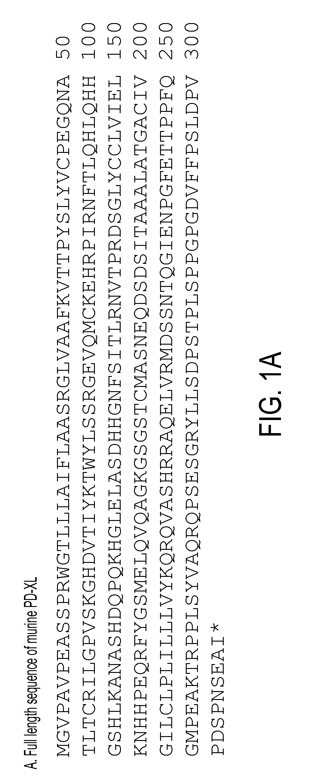
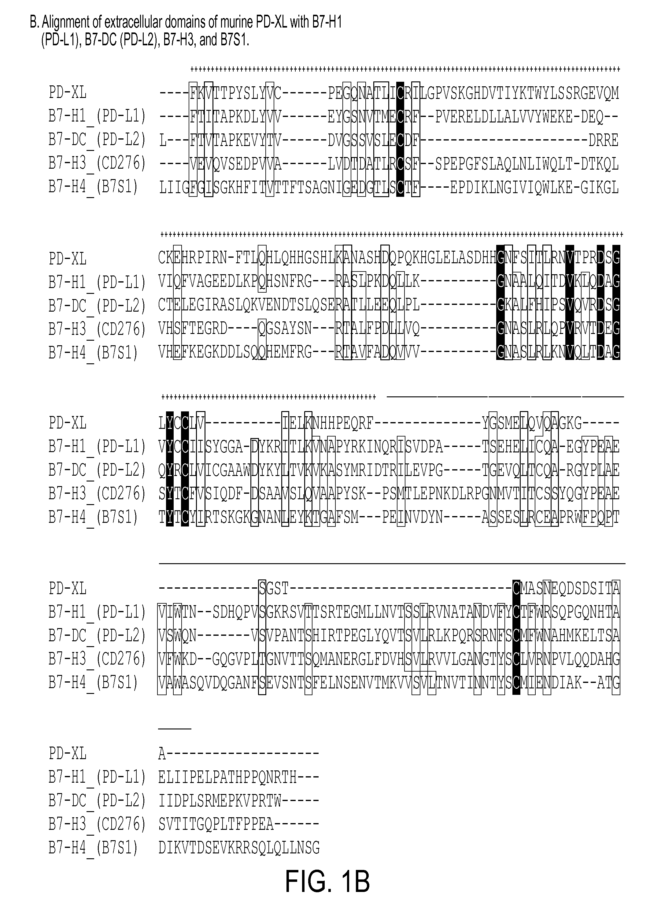
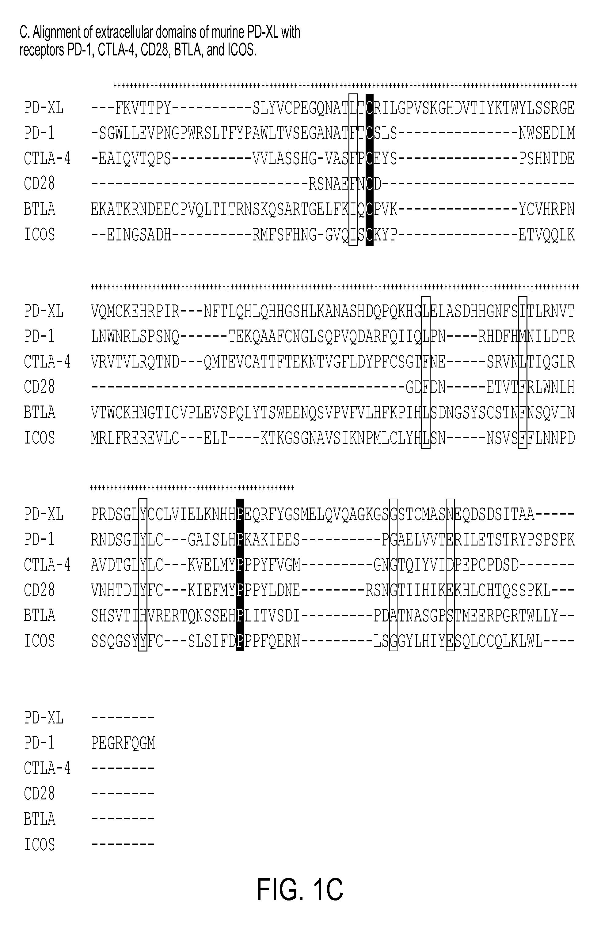
[0304]As shown by the results in FIG. 1A-1D Affymetrix analysis of ac...

example 2

Expression Studies of PD-L3 by RT-PCR Analysis and Flow Cytometry

[0309]As shown in the experiments in FIG. 3, RT-PCR analysis was used to determine the mRNA expression pattern of PD-L3 in mouse tissues (FIG. 3A). PD-L3 is mostly expressed on hematopoietic tissues (spleen, thymus, bone marrow), or tissues with ample infiltration of leukocytes (i.e. lung). Weak expression was also detected in non-hematopoietic tissues (i.e. heart, kidney, brain, and ovary). Analysis of several hematopoietic cell types reveals expression of PD-L3 on peritoneal macrophages, splenic CD11b+ monocytes, CD11c+ DCs, CD4+ T cells and CD8+ T cells, but lower expression level on B cells (FIG. 3B). This expression pattern is also largely consistent with the GNF (Genomics Institute of Novartis Research Foundation) gene array database Su et al., (2002), Proc Natl Acad Sci USA 99, 4465-4470, as well as NCBI GEO (gene expression omnibus) database (FIG. 4).

[0310]In order to study the protein expression, PD-L3 specifi...

example 3

Functional Impact of PD-L3 Signaling on CD4+ and CD8+ T Cell Responses

[0317]A PD-L3-Ig fusion proteins were was produced to examine the regulatory roles of PD-L3 on CD4+ T cell responses. The PD-L3-Ig fusion protein contains the extracellular domain of PD-L3 fused to the human IgG1 Fc region. When immobilized on the microplate, PD-L3-Ig but not control Ig suppressed the proliferation of bulk purified CD4+ and CD8+ T cells in response to plate-bound anti-CD3 stimulation, as determined by arrested cell division (FIG. 9A-B). The PD-L31 g fusion protein did not affect the absorption of anti-CD3 antibody to the plastic wells, as determined by ELISA (data not shown), thus excluding the possibility of non-specific inhibitory effects. PD-1 KO CD4+ T cells were also suppressed (FIG. 9C), indicating that PD-1 is not the receptor for PD-L3. The inhibitory effect of PD-L1-Ig and PD-L3-Ig was also directly compared (FIG. 10). When titrated amounts of Ig fusion proteins were absorbed to the micro...


PUM

PropertyMeasurementUnit
Cell proliferation rateaaaaaaaaaa

Abstract

The present invention relates to novel regulatory T cell proteins. One protein, designated PD-L3, resembles members of the PD-L1 family, and co-stimulates αCD3 proliferation of T cells in vitro. A second, TNF-like, protein has also been identified as being upregulated upon αCD3/αGITR stimulation. This protein has been designated Treg-sTNF. Proteins, antibodies, activated T cells and methods for using the same are disclosed.
In particular methods of using these proteins and compounds, preferably antibodies, which bind or modulate (agonize or antagonize) the activity of these proteins, as immune modulators and for the treatment of cancer, autoimmune disease, allergy, infection and inflammatory conditions, e.g. multiple sclerosis is disclosed

Description

INTRODUCTION[0001]This application is a continuation-in-part of U.S. patent application Ser. No. 11 / 912,397, filed Jun. 4, 2008, which claims benefit of PCT / US2006 / 015239, filed Apr. 24, 2006 and U.S. Provisional Patent Application Ser. No. 60 / 674,567, filed Apr. 25, 2005, the contents of which including the sequence listing are incorporated herein by reference in their entireties.BACKGROUND OF THE INVENTION[0002]Induction of an immune response requires T cell expansion, differentiation, contraction and establishment of T cell memory. T cells must encounter antigen presenting cells (APCs) and communicate via T cell receptor (TCR) / major histocompatibility complex (MHC) interactions on APCs. Once the TCR / MHC interaction is established, other sets of receptor-ligand contacts between the T cell and the APC are required, i.e. co-stimulation via CD154 / CD40 and CD28 / B7.1-B7.2. The synergy between these contacts is suggested to result, in vivo, in a productive immune response capable of cle...

Claims


Application Information

Patent Timeline
IPC IPC(8): A61K39/395A61K38/16C07K16/00C12N5/02C40B30/04
CPCA61K38/00A61K2039/505C07K14/70596C07K2319/30G01N33/505C07K16/2827Y02A50/30C07K2319/00
Inventor NOELLE, RANDOLPH J.WANG, LI
Owner TRUSTEES OF DARTMOUTH COLLEGE THE
Why Eureka
Try Eureka