Devices, systems, and methods to precondition, arterialize and/or occlude a mammalian luminal organ

a luminal organ and device technology, applied in the field of devices, systems, and methods to precondition, arterialize and/or occlude a mammalian luminal organ, can solve the problems of reducing the function of peripheral limbs, reducing the possibility of cardiac or other procedures, and reducing the risk of potential death of patients, etc., to achieve the effect of facilitating localized arterialization, facilitating preconditioning of veins, and increasing fluid pressur

Inactive Publication Date: 2016-07-21
CVDEVICES
View PDF2 Cites 1 Cited by
  • Summary
  • Abstract
  • Description
  • Claims
  • Application Information

AI Technical Summary

Benefits of technology

[0004]In an exemplary embodiment of a device for preconditioning, arterializing, and / or occluding a mammalian luminal organ of the present disclosure, the device comprises a frame comprising a plurality of struts, wherein at least one strut of the plurality of struts forms a local general perimeter or boundary of the device, and an expandable occluder coupled to the frame and comprising a membrane or other expandable material that can generally expand and / or unfold as device shifts from a first configuration to a second configuration. In another embodiment, when the device is deployed within a luminal organ, the device can remain in place within the luminal organ for as long as desired. In yet another embodiment, the first configuration is a generally compressed configuration, and wherein the second configuration is a generally expanded or deployed configuration. In an additional embodiment, the first configuration allows the device to be delivered intravascularly within a patient. In yet an additional embodiment, the expandable occluder is located at or near a distal end of the device.
[0005]In an exemplary embodiment of a device for preconditioning, arterializing, and / or occluding a mammalian luminal organ of the present disclosure, the expandable occluder is located at a relative center / middle of the device. In an additional embodiment, when the device is deployed within a luminal organ, the expandable occluder hinders blood flow through the luminal organ at the location of the expandable occluder. In yet an additional embodiment, the expandable occluder comprises a material selected from the group consisting of a biological material and a biologically-compatible material, but not limited to, polytetrafluoroethylene (PTFE), visceral pleura, lung ligament tissue, and other suitable bodily (mammalian) tissues. In another embodiment, the expandable occluder is connected to the frame using one or more sutures. In yet another embodiment, the plurality of struts includes at least one perimeter strut and / or at least one lateral strut used, for example, to minimize blockage of side branches.
[0009]In an exemplary embodiment of a method of using an exemplary device of the present disclosure, the method comprises the steps of inserting an exemplary device of the present disclosure within a non-arterial luminal organ, the exemplary device comprising a frame and an expandable occluder coupled thereto, expanding the exemplary device from a first, collapsed configuration to a second, expanded configuration to anchor the exemplary device within the luminal organ, wherein the exemplary device, when in the second, expanded configuration, is operable to block bodily fluid therethrough at the location of the expandable occluder. In another embodiment, the exemplary device, when expanded and anchored in a vein, facilitates preconditioning of the vein prior to arterialization. In yet another embodiment, the exemplary device, when expanded and anchored in a vein, facilitates localized arterialization at or near the expandable occluder. In an additional embodiment, when the exemplary device is expanded and anchored in a vein at a bifurcation having a first side branch and a second side branch, blood can flow through the first side branch but not the second side branch.
[0010]In an exemplary embodiment of a method of using an exemplary device of the present disclosure, the exemplary device, when expanded and anchored in the luminal organ, causes a localized stenosis at or near the expandable occluder. In another embodiment, the step of expanding is performed to cause an occlusion within the non-arterial luminal organ due to expansion of the expandable occluder. In an additional embodiment, the exemplary device, when expanded and anchored in the luminal organ, causes a thrombosis at or near the expandable occluder. In yet an additional embodiment, the exemplary device, when expanded and anchored in the luminal organ, causes an increase in fluid pressure within the luminal organ at or near the expandable occluder. In another embodiment, the exemplary device, when expanded and anchored in the luminal organ, facilitates thickening of walls (considered as functional arterialization) of the luminal organ at or near (such as distal to) the expandable occluder.
[0011]In an exemplary embodiment of a method of using an exemplary device of the present disclosure, the exemplary device, when expanded and anchored in an opening of an atrial appendage, prevents blood flow in and / or out of the atrial appendage, such as for atrial fibrillation patients that are prone to thrombus generation in the appendage. In an additional embodiment, the step of inserting is performed to position the device within a vein, and wherein the device, when in the second, expanded configuration, blocks blood flow through the vein at the expandable occluder. In yet an additional embodiment, the device is configured to fully block a flow of bodily fluid in the non-arterial luminal organ at the expandable occluder. In another embodiment, the method further comprises the step of performing a medical procedure after the exemplary device is anchored within the luminal organ. In yet another embodiment, the medical procedure comprises a coronary bypass graft procedure or an anastomosis procedure. In various embodiments, the medical procedure is performed to treat a peripheral circulatory system condition. In at least one embodiment, the plurality of struts includes at least two perimeter struts and at least one lateral strut, the at least one lateral strut connected to two of the at least two perimeter struts, and wherein the step of inserting is performed to insert the device within a non-arterial luminal organ at a bifurcation so that the bifurcation is located at the at least one lateral strut.

Problems solved by technology

Thousands, if not millions, of patients in the U.S. and across the globe suffer from cardiac, peripheral, and other circulatory conditions to such an extent that further cardiac or other procedures are no longer possible with current medical technology.
As such, said patients are at the risk of potential death, loss of peripheral limb function or limbs altogether, or other catastrophic or potentially catastrophic conditions, due to diseases and other conditions within their circulatory systems.

Method used


Image

  • Devices, systems, and methods to precondition, arterialize and/or occlude a mammalian luminal organ
  • Devices, systems, and methods to precondition, arterialize and/or occlude a mammalian luminal organ
  • Devices, systems, and methods to precondition, arterialize and/or occlude a mammalian luminal organ

Examples


Embodiment Construction

[0021]For the purposes of promoting an understanding of the principles of the present disclosure, reference will now be made to the embodiments illustrated in the drawings, and specific language will be used to describe the same. It will nevertheless be understood that no limitation of the scope of this disclosure is thereby intended.

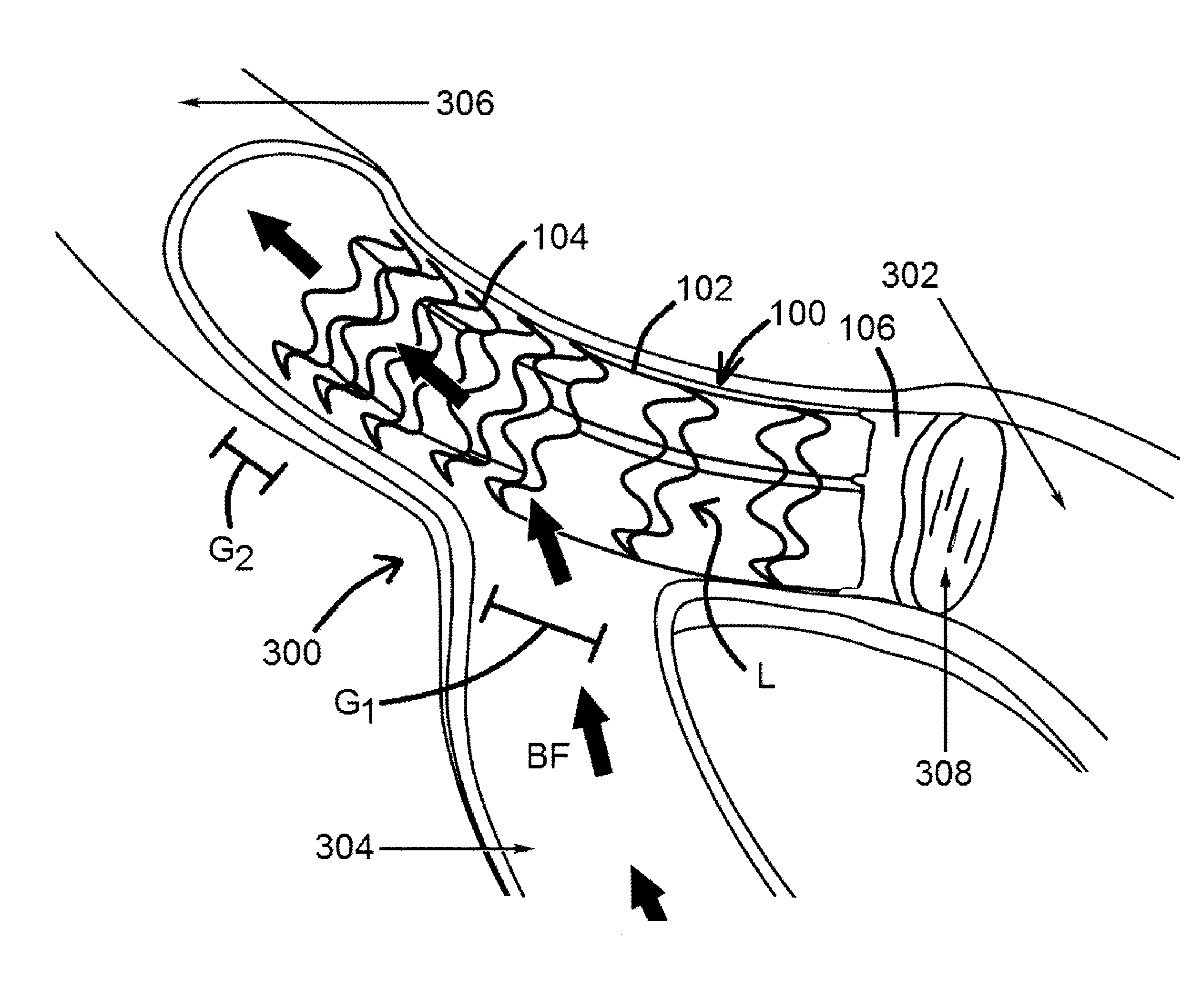
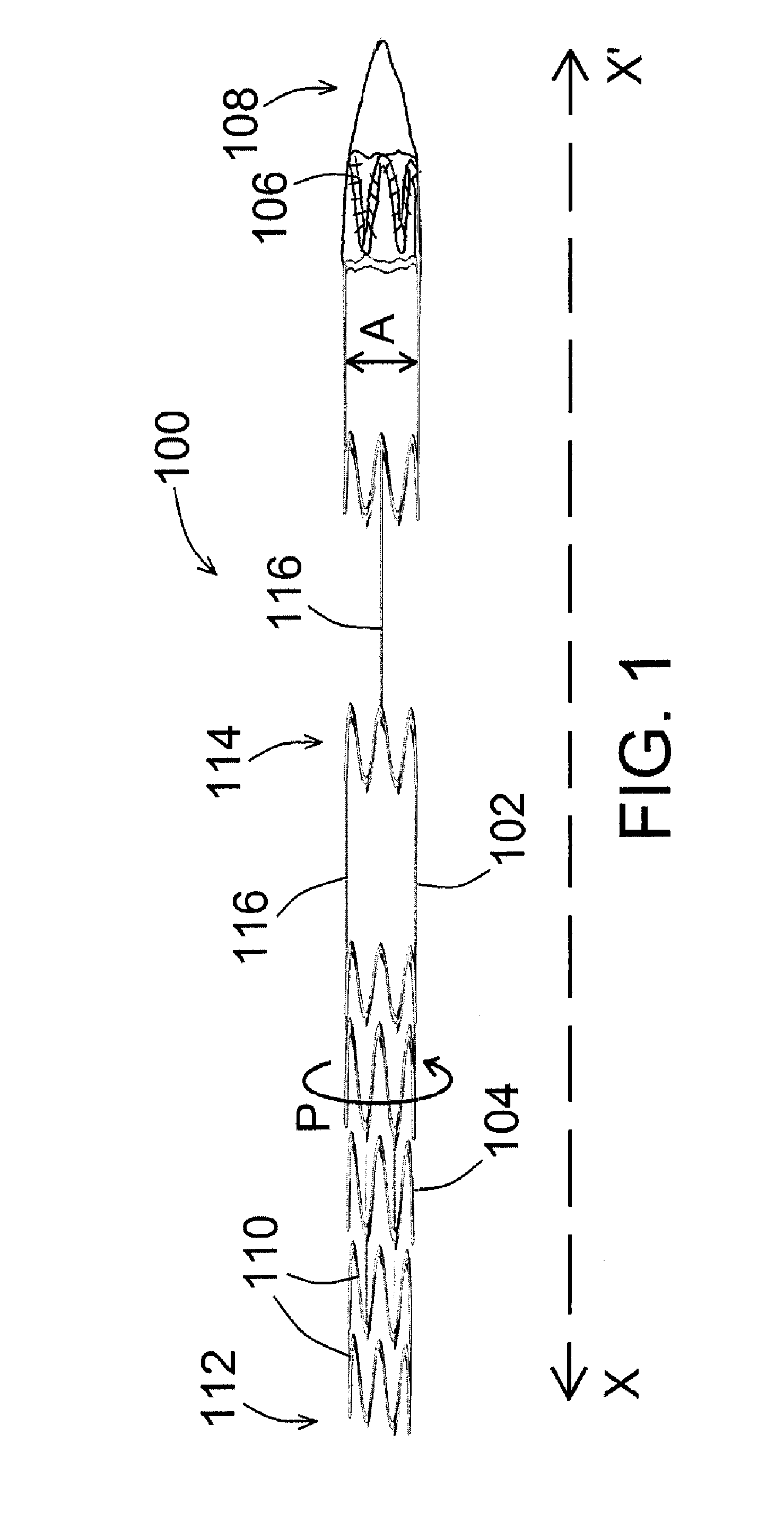
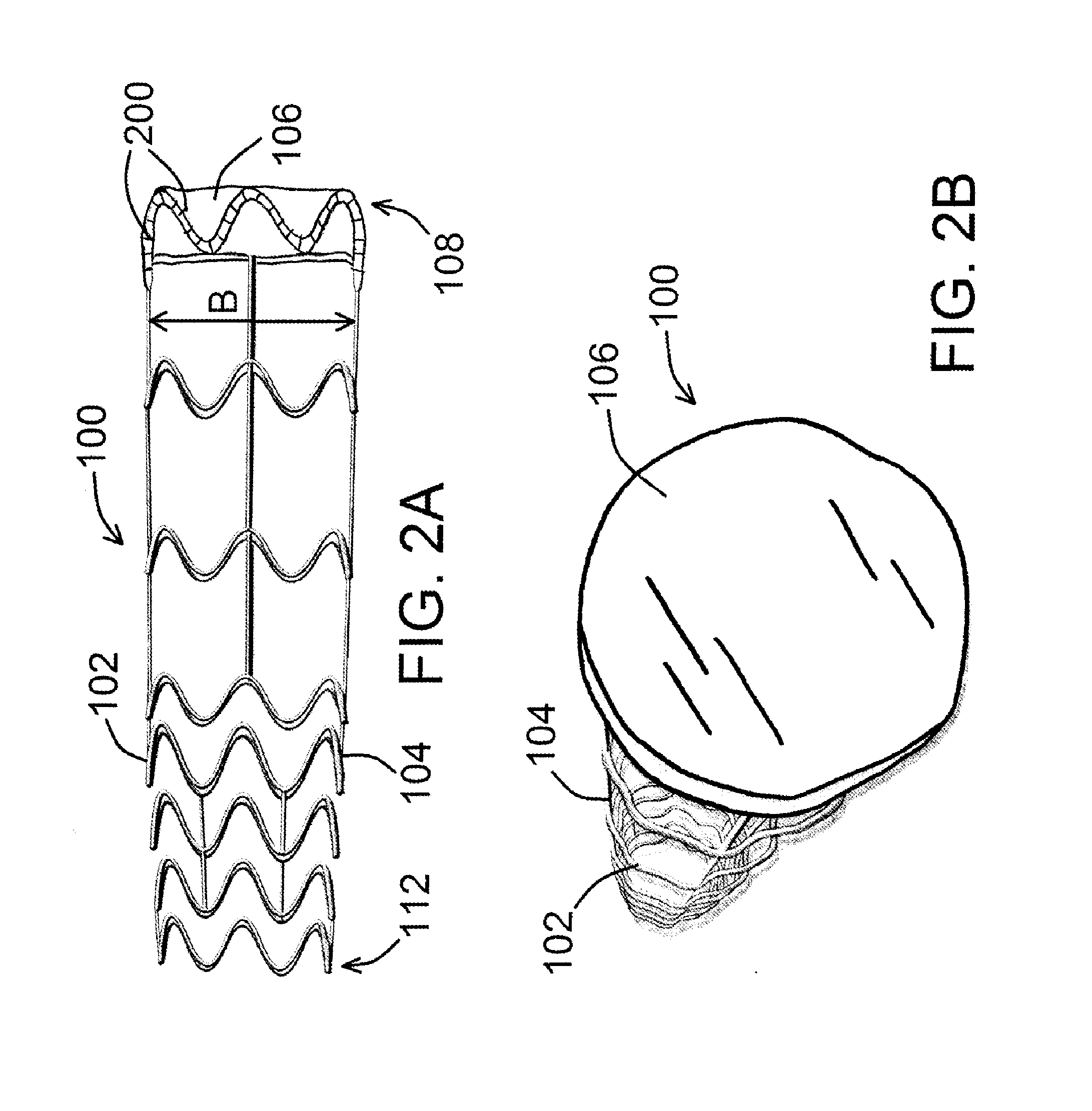
[0022]An exemplary device for preconditioning and / or occluding a luminal organ of the present disclosure is shown in FIG. 1. As shown in FIG. 1, device 100 comprises a frame 102 having a number of struts 104, whereby at least one of the various struts 104 forms a local general perimeter or boundary of device 100 (shown as P in the figure) so that device 100, when ultimately deployed within a luminal organ 300 (such as a blood vessel, for example, as shown in FIG. 3), device 100 can remain in place within said luminal organ 300 for as long as desired. Device 100 is shown in a generally compressed configuration in FIG. 1, and is shown in a generally expan...


PUM


Abstract

Devices, systems, and methods to precondition, arterialize, and / or occlude a mammalian luminal organ. In an exemplary embodiment of a device for preconditioning, arterializing, and / or occluding a mammalian luminal organ of the present disclosure, the device comprises a frame comprising a plurality of struts, wherein at least one strut of the plurality of struts forms a local general perimeter or boundary of the device, and an expandable occluder coupled to the frame and comprising a membrane or other expandable material that can generally expand and / or unfold as device shifts from a first configuration to a second configuration.

Description

PRIORITY[0001]The present international patent application is related to, and claims the priority benefit of, U.S. Provisional Patent Application Ser. No. 61 / 882,837, filed Sep. 26, 2013, the contents of which are hereby incorporated by reference in their entirety into the present disclosure.BACKGROUND[0002]Thousands, if not millions, of patients in the U.S. and across the globe suffer from cardiac, peripheral, and other circulatory conditions to such an extent that further cardiac or other procedures are no longer possible with current medical technology. As such, said patients are at the risk of potential death, loss of peripheral limb function or limbs altogether, or other catastrophic or potentially catastrophic conditions, due to diseases and other conditions within their circulatory systems.[0003]In view of the same, devices and systems useful to precondition, arterialize, and / or occlude a mammalian luminal organ, to address at least the conditions identified above, would be w...

Claims


Application Information

Patent Timeline
Patent Type & Authority Applications(United States)
IPC IPC(8): A61B17/12
CPCA61B17/12172A61B17/12109A61B2017/00292A61B17/1204A61B17/12122A61B17/12031A61B17/12177
Inventor KASSAB, GHASSAN S.
Owner CVDEVICES
Why Eureka
Try Eureka