

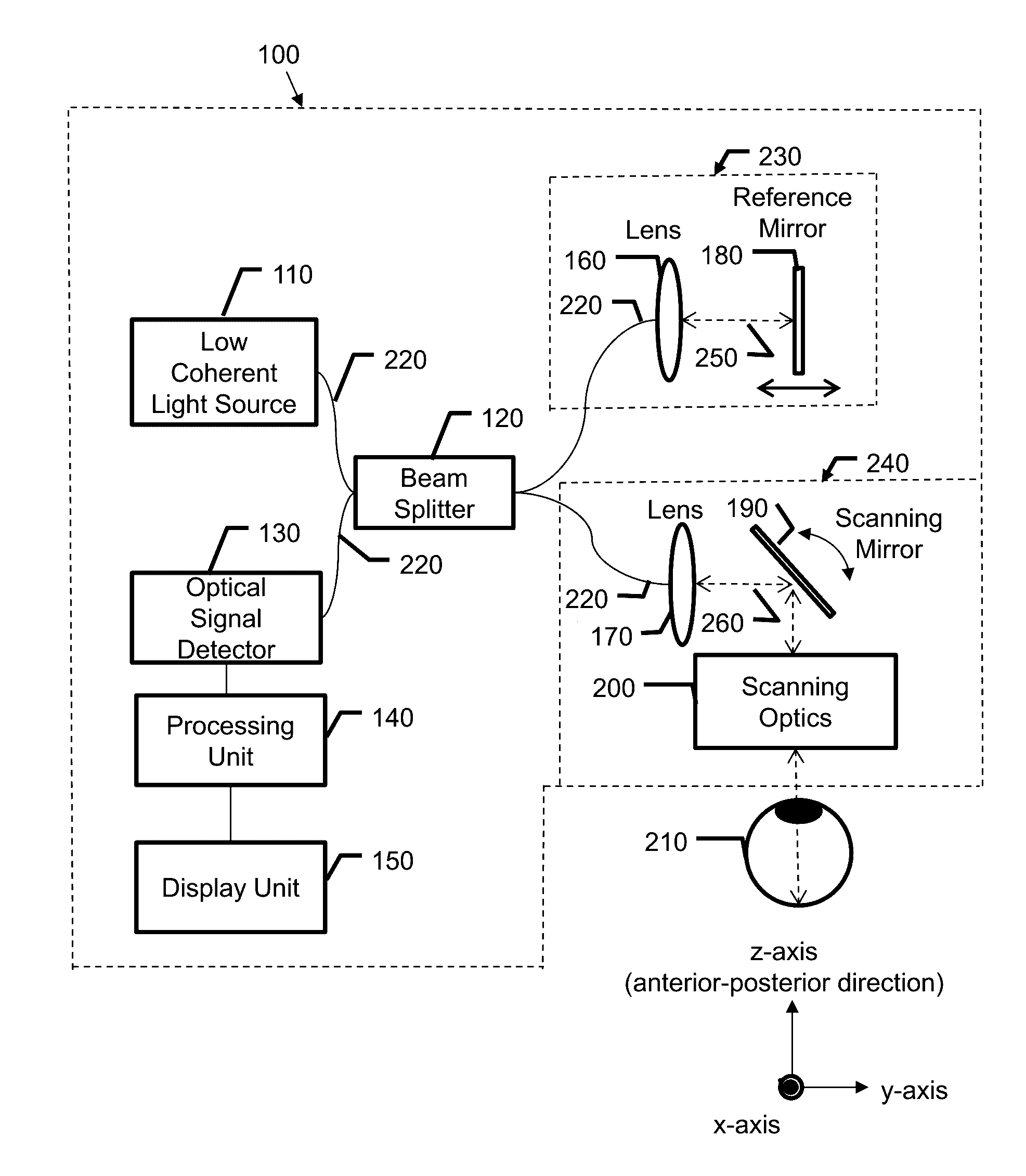
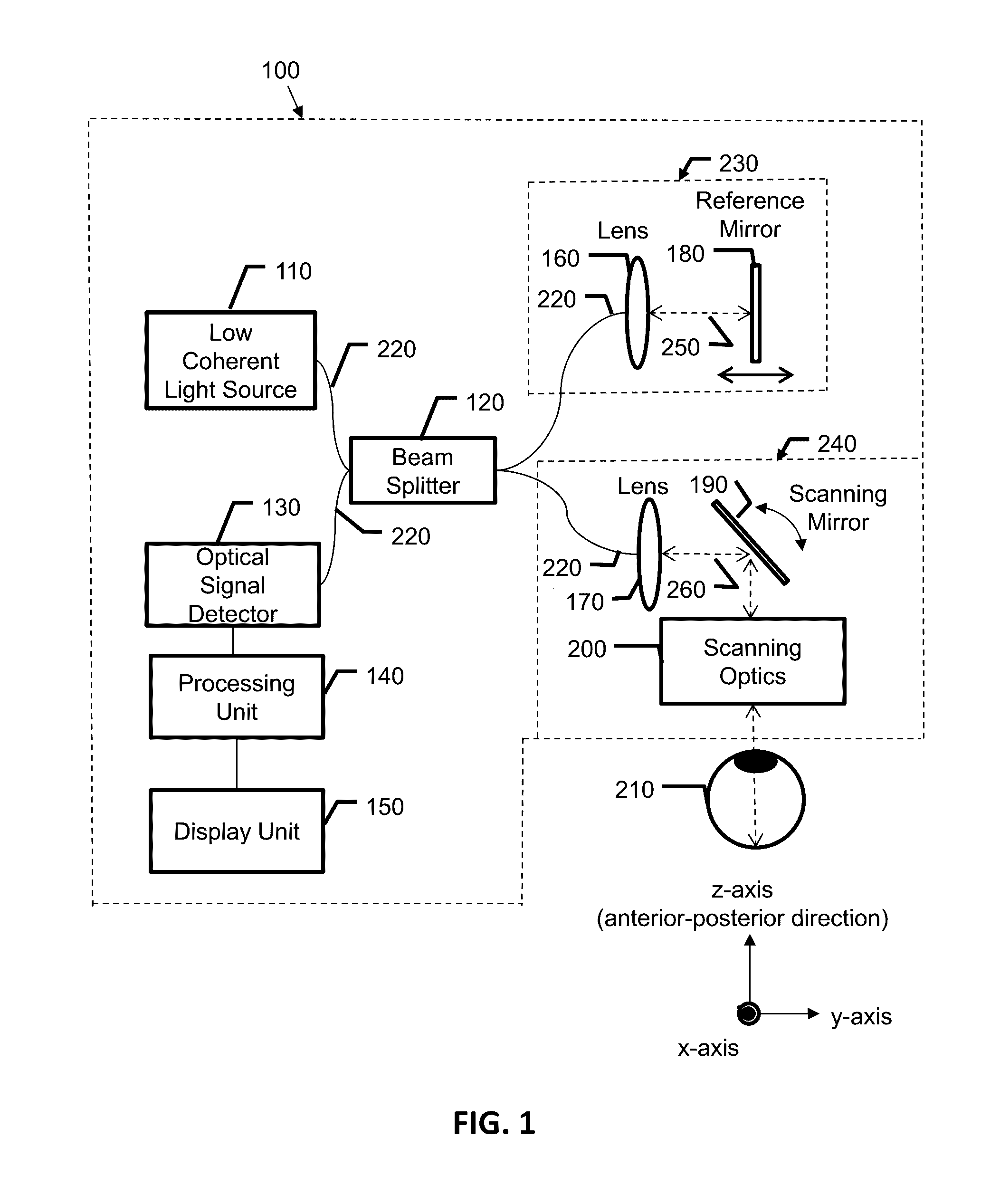
Optical coherence tomography system for health characterization of an eye
a coherence tomography and optical coherence technology, applied in the field of optical coherence tomography, can solve the problems of inconvenient use, inconvenient use, and inability to accurately depict the fine anatomic structure of the choriocapillaries
- Summary
- Abstract
- Description
- Claims
- Application Information
AI Technical Summary
Benefits of technology
Problems solved by technology
Method used
Image

Examples
example 1
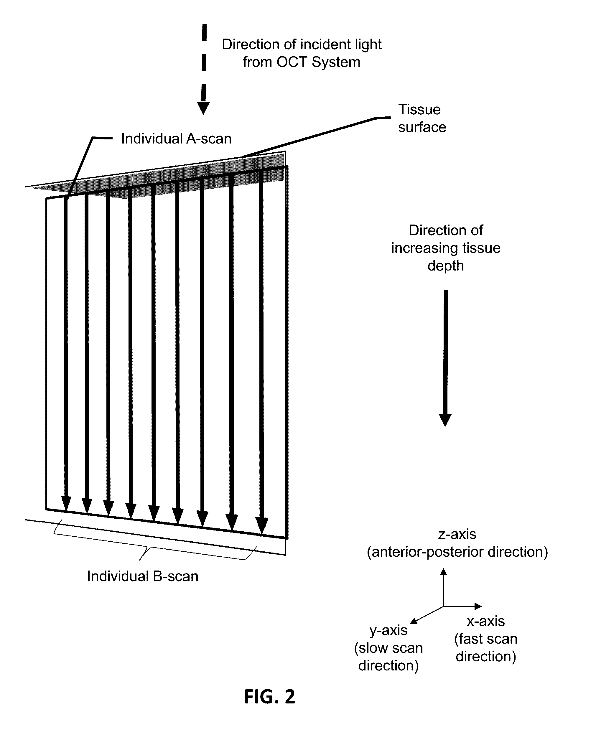
[0110]High resolution OCT volumetric scans may be acquired over the retina. En face images from several different retinal layers may be extracted and the vasculature may be isolated from these layers, and the two dimensional vascular density maps may be calculated. These vascular density maps may spatially vary across the retina, typically going to zero in the foveal avascular zone. The vascular density maps from the retinal layers may be aggregated into a single quantitative number to characterize the vascular health of the retina.
example 2
[0111]Wide field high resolution cross-sectional OCT angiography scans may be acquired at sparsely sampled locations across the retina. For example, for a 20×20 degree field of view, OCT angiography images may be acquired at 5° separations to allow for quicker, wide field acquisitions. Microvascular projections may be identified within the cross-sectional scans, and vascular densities may be calculated along the horizontal direction of the scan. These calculations may be repeated for the other cross-sectional images, giving a 2D vascular density map across a wide field of the retina, with low resolution along the slow axis direction.
example 3
[0112]Wide field cross-sectional OCT angiography scans may be acquired, but the entire horizontal range may be broken up into several, smaller acquisitions which may reduce the phase noise errors present. These acquisitions may be stitched together in the horizontal direction to cover a wider range. Each of these acquisitions may also be acquired as a small volumetric scan in the slow scanning axis directly, only about 0.5° to provide some additional context and statistics to identify the microvasculature within the retina. These scans are also repeated sparsely over the slow axis direction to cover a wider field of view.
[0113]The OCT system disclosed above may be used for any OCT related application. For example, this system maybe used in forming larger field of view OCT images of the physical object. This system may be incorporated into methods and systems related to OCT based angiography. For example, the choroidal vasculature may be identified in more detail by using the OCT sys...
PUM
 Abstract
Description
Claims
Application Information