System and method for three-dimensional quantitative evaluaiton of uterine fibroids

a three-dimensional quantitative and evaluaiton technology, applied in image enhancement, tomography, instruments, etc., can solve the problems of uterine fibroids, heavy menstrual bleeding, and -related symptoms

Inactive Publication Date: 2016-11-17
KONINKLJIJKE PHILIPS NV
View PDF5 Cites 10 Cited by
  • Summary
  • Abstract
  • Description
  • Claims
  • Application Information

AI Technical Summary

Problems solved by technology

Clinically, uterine fibroids can lead to heavy menstrual bleeding as well as bulk-related symptoms such as severe lower abdominal pain and in some patients even infertility.
However, these techniques lead to reader bias and imprecision.
In addition to lacking reliability, the measurements are difficult to reproduce.
Numerous studies have failed to achieve a reliable correlation between imaging results and clinical symptom relief.
Accordingly, the studies have failed to establish contrast-enhanced MRI as a suitable tool for evaluating fibroid response to UAE or to detect insufficiently treated patients.
The correlation of late follow-up MRI results with clinical symptoms using a relative approach may be inaccurate.

Method used


Image

  • System and method for three-dimensional quantitative evaluaiton of uterine fibroids
  • System and method for three-dimensional quantitative evaluaiton of uterine fibroids
  • System and method for three-dimensional quantitative evaluaiton of uterine fibroids

Examples


Embodiment Construction

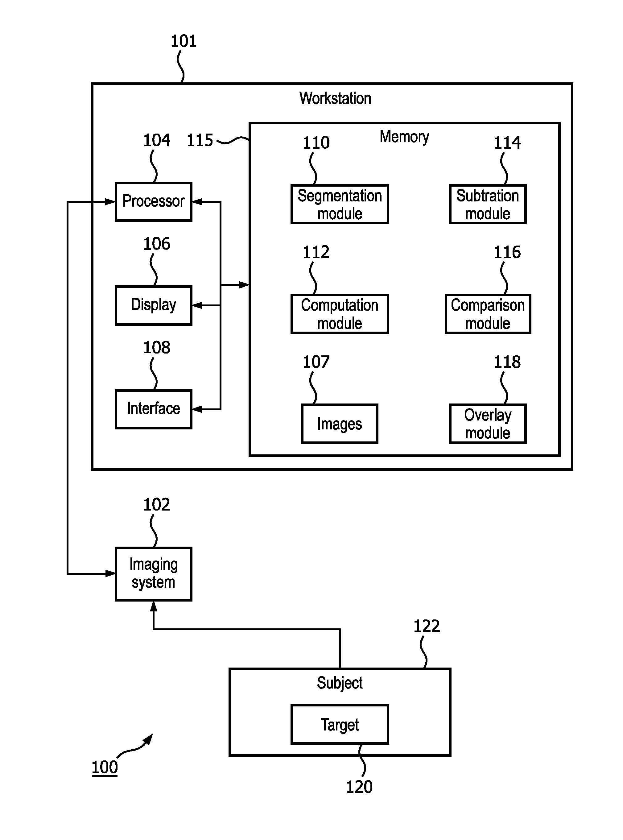
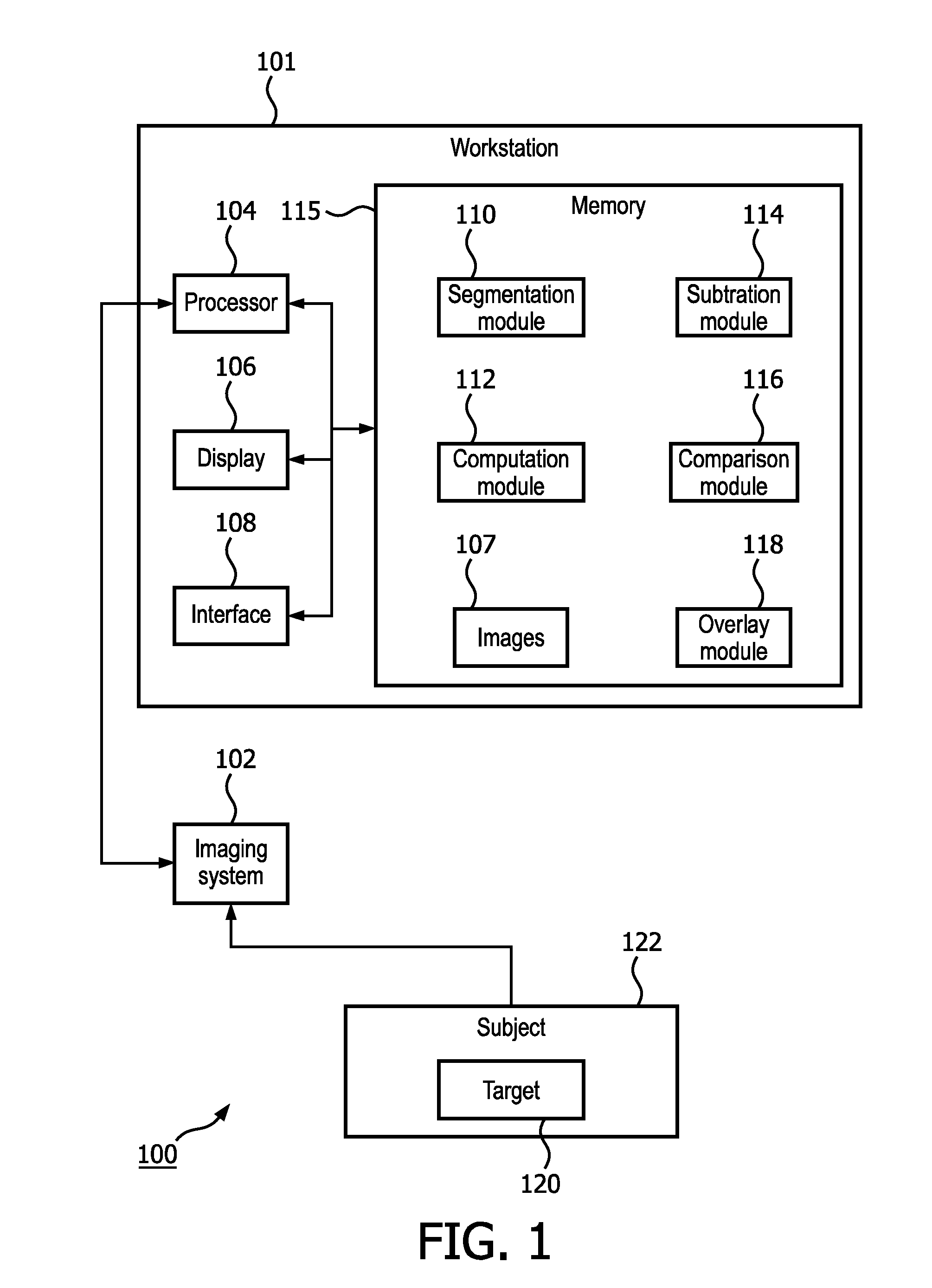
[0021]In accordance with the present principles, a three-dimensional quantitative evaluation of uterine fibroids is performed in which absolute quantification of the overall fibroid volume and the enhancing fibroid volume is performed. The system and method provide a reproducible and accurate quantification of the viable total fibroid volume and enhancing fibroid volume in order to measure uterine fibroid enhancement.

[0022]The system performs a three-dimensional segmentation of a fibroid lesion on contrast-enhanced arterial phase MRI or cone-beam computed tomography (“CBCT”) images. The total volume of the lesion is computed from the three-dimensional segmentation. A differentiation process is then employed which distinguishes between actual contrast enhancement and background or baseline enhancement. Background or baseline enhancements include false positive enhancements in the image as well as noise. The three-dimensional segmentation mask is then applied to the differentiated ima...


PUM


Abstract

A system (100) and method which assess the results of uterine fibroid embolization. A fibroid lesion is three-dimensionally segmented by a segmentation module (110) on contrast-enhanced arterial phase MRI or CBCT images and a differentiation process performed by a subtraction module (114) identifies actual contrast enhancement from background enhancement. The three-dimensional segmentation mask is then applied to the differentiated image and a region of interest is defined by a comparison module (116) in order to define a normalized threshold. A computation module (112) performs a voxel-by-voxel analysis of enhancing fibroid volume and quantifies the viable enhanced fibroid volume and overall fibroid volume in absolute numerical figures.

Description

RELATED U.S. APPLICATION[0001]This application claims the benefit of U.S. Patent Application Ser. No. 61 / 930,989 filed on Jan. 24, 2014 and 62 / 022,695, filed on Jul. 10, 2014.[0002]This invention was made with government support under grant no. R01 CA160771-01 awarded by the National Cancer Institute of the United States National Institutes of Health. The government has certain rights in the invention.BACKGROUND[0003]1. Technical Field[0004]This disclosure relates to image guided intervention, therapy and evaluation, in particular, to an apparatus and method for obtaining reproducible, three-dimensional quantitative assessment of uterine fibroids before, during and after inter-arterial therapy.[0005]2. Description of the Related Art[0006]Uterine fibroids are the most common benign tumors in females, typically diagnosed in the middle reproductive years. Clinically, uterine fibroids can lead to heavy menstrual bleeding as well as bulk-related symptoms such as severe lower abdominal pa...

Claims


Application Information

Patent Timeline
Patent Type & Authority Applications(United States)
IPC IPC(8): G06T7/00A61B6/03A61B5/026A61B6/00
CPCG06T7/0014A61B6/481A61B6/4085A61B6/03A61B6/486G06T2207/10028A61B5/0263A61B6/5217G06T7/0081G06T2207/10096G06T2207/30004A61B6/507A61B5/055G06T7/11G06T7/62G16H50/30
Inventor LIN, MING DECHAPIRO, JULIUSHONG, KELVIN KEIVIN
Owner KONINKLJIJKE PHILIPS NV
Why Eureka
Try Eureka