Imaging diagnosis apparatus having needling navigation control system and a needling navigation controlling method

a technology of needling navigation control and imaging diagnosis, which is applied in the field of imaging diagnosis apparatus and needling navigation control method, can solve the problems of difficult to accurately insert the puncturing needle into the tumor portion without injuring the surrounding tissues of the tumor portion such as blood vessels and other organs, and the conventional techniques that are proposed cannot confirm the inserting status of the puncturing needle on the 2-d image data

Active Publication Date: 2017-04-27
CANON MEDICAL SYST COPRPORATION
View PDF9 Cites 10 Cited by
  • Summary
  • Abstract
  • Description
  • Claims
  • Application Information

AI Technical Summary

Problems solved by technology

However, during insertion of the puncturing needle, it is usually happened to shift the inserting direction of the puncturing needle from a prescribed slice plane for acquiring image data due to unevenness of living body tissues existing along the insertion route, i.e., differences among muscle portions and fatty portions.
In such a case, the proposed conventional techniques could not confirm the inserting status of the puncturing needle on the 2-D image data.
Consequently, it has difficult to accurately insert the puncturing needle into the tumor portion without injuring the surrounding tissues of the tumor portion such as the blood vessels and other organs.

Method used


Image

  • Imaging diagnosis apparatus having needling navigation control system and a needling navigation controlling method
  • Imaging diagnosis apparatus having needling navigation control system and a needling navigation controlling method
  • Imaging diagnosis apparatus having needling navigation control system and a needling navigation controlling method

Examples


Embodiment Construction

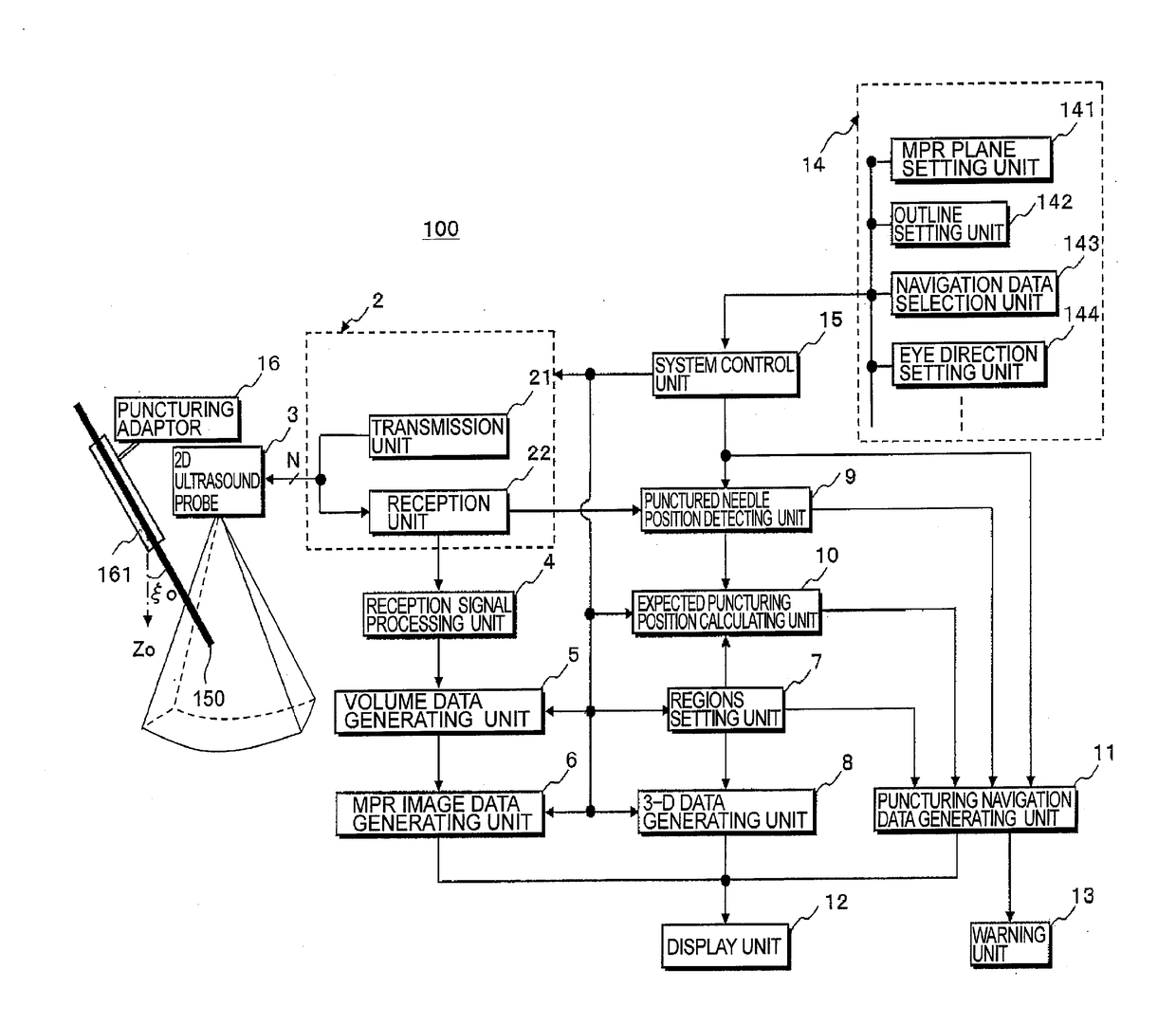
[0046]In the preferred embodiment consistent with the present invention, a target tumor region for puncturing (hereinafter, “tumor region”) and, major blood vessel regions (hereinafter, “blood vessel regions”) and other organ region located near to the tumor region (hereinafter, “organ region”) are set based on the volume data acquired by the 3-D scan on the object in order to avoid any possibility of wrong insertion by the puncturing needle into the blood vessel regions and the organ region located near the tumor region.

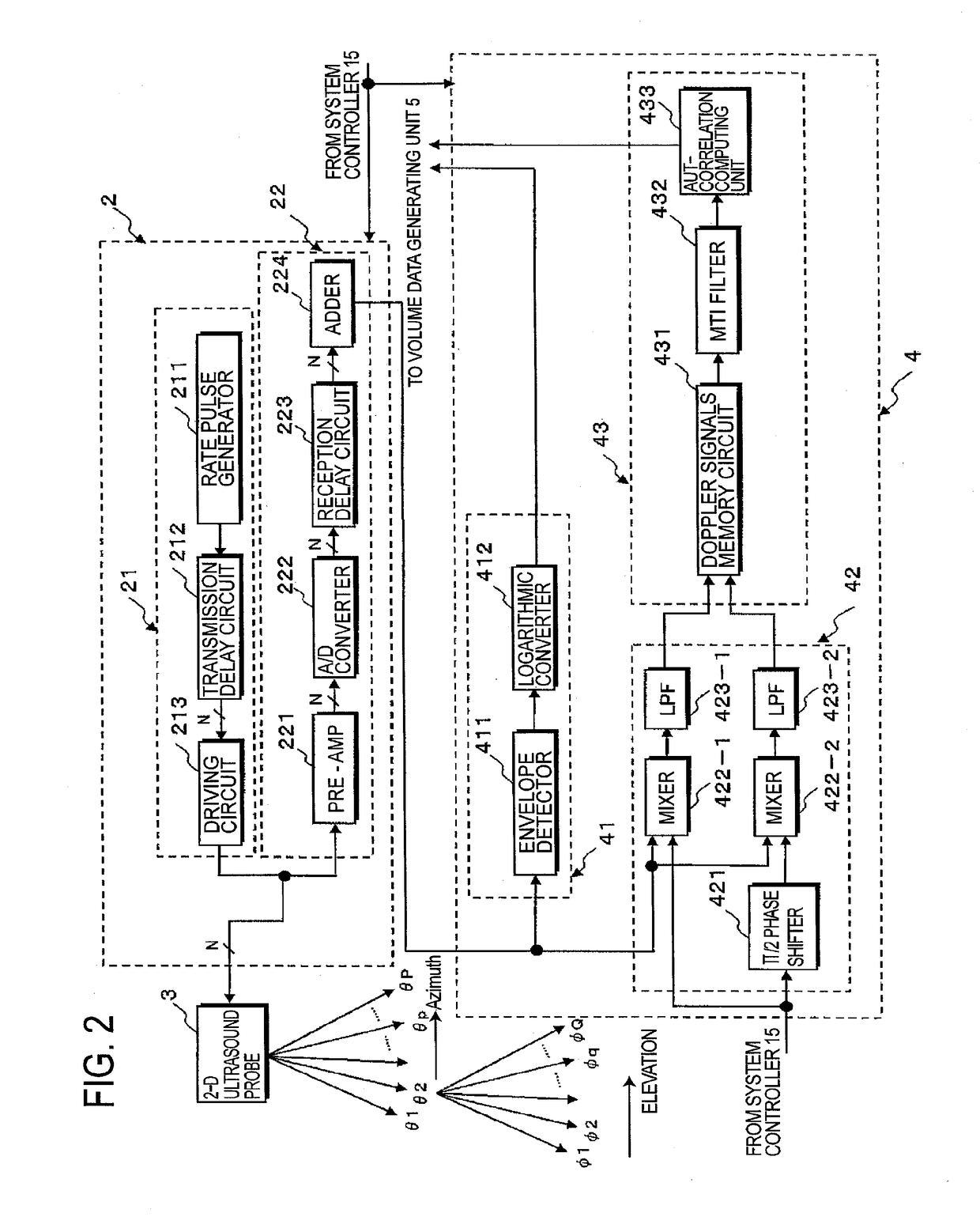
[0047]In the preferred embodiments of the ultrasound imaging diagnosis apparatus consistent with the present invention, based on 3-D B mode data and 3-D color Doppler data acquired by a 2-D array ultrasound probe in which a plurality of transducers are two-dimensionally arranged, volume data are generated. By using the volume data of the 3-D B mode data, the tumor region and the organ region are approximately set as a sphere or an ellipse solid. By using volume data...


PUM


Abstract

An ultrasound imaging diagnosis apparatus and a method for supporting safe and exact puncturing into a target region in a patient's body are provided. In the apparatus, a regions setting unit sets a target tumor region for puncturing, blood vessel and organ regions that are located near the tumor region based on 3-D volume data. To avoid insertions into blood vessel and organ regions, a needle position detecting unit detects the tip position and inserting direction of the needle before and during insertion. An expected inserting position calculating unit calculates an expected inserting position and an insertion error region to the tumor region based on the tip position data, inserting direction data and needle characteristic data. A puncturing navigation data generating unit generates puncturing navigation data by composing tumor region data, organ region and blood vessel regions data, and data relating to the expected inserting position and insertion error region.

Description

CROSS-REFERENCE TO RELATED APPLICATIONS[0001]This application is a continuation of and is based upon and claims the benefit under 35 U.S.C. §120 for U.S. application Ser. No. 12 / 275,886, filed Nov. 21, 2008, and claims the benefit of priority from, under 35 U.S.C. §119 from Japanese Patent Application No. 2007-303040, filed on Nov. 22, 2007. The contents of which are incorporated herein by reference.BACKGROUND OF THE INVENTION[0002]A. Field of the Invention[0003]The present invention relates to an imaging diagnosis apparatus, such as an ultrasound diagnosis apparatus and a computer tomography (CT) apparatus, having a needling navigation control system and a needling navigation controlling method. More particularly, the present invention relates to an imaging diagnosis apparatus and a needling navigation controlling method that can accurately and safely navigate an invasive instrument, such as a puncturing needle, into a diagnostic target organ in the body of an object with monitorin...

Claims


Application Information

Patent Timeline
Patent Type & Authority Applications(United States)
IPC IPC(8): A61B8/08A61B34/20A61B8/00
CPCA61B8/0841A61B8/483A61B34/20A61B8/466A61B8/5207A61B6/12A61B8/0833
Inventor TAKIMOTO, MASAOSAKAGUCHI, FUMIYASU
Owner CANON MEDICAL SYST COPRPORATION
Why Eureka
Try Eureka