Unlock instant, AI-driven research and patent intelligence for your innovation.

Anterior eye three-dimensional image processing apparatus and method of anterior eye three-dimensional image processing

Active Publication Date: 2017-05-18
TOMEY CORP
View PDF0 Cites 0 Cited by
  • Summary
  • Abstract
  • Description
  • Claims
  • Application Information

AI Technical Summary

Benefits of technology

The present patent is about an improved anterior eye three-dimensional image processing apparatus and method that can identify specific positions in tomographic images with greater accuracy and stability. This reduces the burden of examiners and helps in diagnosing and treating eye-related issues. The apparatus uses a processor and memory to acquire multiple tomographic images of the eye and identify temporary positions. It then calculates a reference circle based on these positions and identifies additional temporary positions in non-representative images. The temporary positions are then corrected based on edge lines indicating tissue boundaries. This allows for accurate identification of scleral spurr positions and faster processing.

Problems solved by technology

Therefore, there was a problem that even if the anterior eye OCT could obtain more than a hundred two-dimensional tomographic images, a large amount of time was taken until a start of creating the chart showing the ITC such that it was difficult to use the apparatus in clinical practice.

Method used


Image

  • Anterior eye three-dimensional image processing apparatus and method of anterior eye three-dimensional image processing
  • Anterior eye three-dimensional image processing apparatus and method of anterior eye three-dimensional image processing
  • Anterior eye three-dimensional image processing apparatus and method of anterior eye three-dimensional image processing

Examples


first embodiment

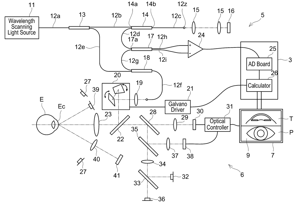
[0045]An anterior eye optical coherence tomographic image capturing apparatus of a first embodiment is used for ophthalmologic examinations for an anterior eye Ec of a subject's eyeball (subjected eye E) (see FIG. 1) such as an angle analysis, and measurements of a corneal curvature, a distribution of corneal thickness, an anterior chamber depth, and the like. The anterior eye optical coherence tomographic image capturing apparatus obtains a three-dimensional (3D) image by capturing two-dimensional (2D) tomographic images of the anterior eye Ec of the subjected eye E by Optical Coherence Tomography (OCT). Hereinbelow, the anterior eye optical coherence tomographic image capturing apparatus is referred to as “anterior eye OCT 1”.

[0046]Although not shown, a main body of the anterior eye OCT 1 is supported so as to be movable in an X direction (left-and-right direction), in a Y direction (up-and-down direction), and in a Z direction (front-and-rear direction) relative to a holding tabl...

second embodiment

[0131]Next, a second embodiment of the present disclosure will be described. Notably, the second embodiment is different from the first embodiment only in its content of the main processing (anterior eye 3D image processing) performed by the image processor 100, and hence descriptions for other contents of the second embodiment will be omitted.

[0132]Specifically, in the anterior eye 3D image processing of the first embodiment, by performing the tomographic image positional displacement adjustment processing (S25), the image processor 100 put together the space coordinate positions of the respective 2D tomographic images, and restructured the anterior eye 3D image, and then identified the SS positions in the respective 2D tomographic images constituting the restructured anterior eye 3D image (S30 to S65).

[0133]Contrary to this, the anterior eye 3D image processing of the second embodiment is different in a point that the image processor 100 determines the SS positions by using parame...


PUM


Abstract

An anterior eye three-dimensional (3D) image processing apparatus performs: identifying first temporary SS positions in each of at least two representative images selected from a plurality of 2D tomographic images constituting an anterior eye 3D image, each first temporary SS position indicating a space coordinate position of a scleral spur of the subjected eye; calculating a reference circle passing through at least three of the first temporary SS positions; identifying second temporary SS positions in each of at least one non-representative image on the calculated reference circle; extracting regions in a predetermined range in each 2D tomographic image, each region being centered at a corresponding one of the identified first or second temporary SS positions; identifying edge lines each of which indicating a tissue boundary that exists in each extracted region; and correcting the identified first or the second temporary SS positions based on the identified edge lines.

Description

CROSS-REFERENCE TO RELATED APPLICATION[0001]This application claims priority to Japanese Patent Application No. 2015-224339 filed on Nov. 17, 2015, the contents of which are hereby incorporated by reference into the present application.TECHNICAL FIELD[0002]The present disclosure relates to an anterior eye three-dimensional image processing apparatus configured to process a three-dimensional image of an anterior eye of a subjected eye and a method thereof.TECHNICAL BACKGROUND[0003]In recent years, as an examination apparatus used for ophthalmologic examination, an optical coherence tomographic image capturing apparatus (hereinbelow referred to as “anterior eye OCT”) which captures a tomographic image of a subject's eyeball (subjected eye) by an optical coherence tomography (OCT) is provided.[0004]Specifically, the anterior eye OCT is used, for example, for glaucomatous examination. A main part of glaucomatous examination is angle analysis of a narrow-angled eye with primary angle clo...

Claims


Application Information

Patent Timeline
IPC IPC(8): A61B3/00A61B3/117A61B3/10
CPCA61B3/0025A61B3/117A61B3/102
Inventor OKAMOTO, KEIICHIROHIGASHITA, RISA
Owner TOMEY CORP