Apparatus and methods for treating excess intraocular fluid

a technology of excess intraocular fluid and apparatus, which is applied in the direction of positive displacement liquid engine, pump parameter, prosthesis, etc., can solve the problems of total blindness, loss of vision, optic nerve, etc., and achieve the effect of avoiding hypotony and minimizing the buildup of proteinaceous sedimen

Active Publication Date: 2017-12-07
ECOLE POLYTECHNIQUE FEDERALE DE LAUSANNE (EPFL)
View PDF0 Cites 4 Cited by
  • Summary
  • Abstract
  • Description
  • Claims
  • Application Information

AI Technical Summary

Benefits of technology

[0021]The present invention overcomes the drawbacks of previously-known ocular drainage systems by providing an implantable device having few moving parts, and which may be non-invasively periodically adjusted to control the fluidic resistance of the device, thereby avoiding hypotony and enabling intraocular pressure to be maintained within desired limits over extended periods. In addition, the ocular drainage device of the present invention is configured so as to minimize buildup of proteinaceous sediment from the aqueous humor on moving parts of the device, thereby ensuring that the implantable device remains functional and adjustable over extended periods.
[0023]In accordance with one aspect of the present invention, an edge of the disk bears against the deformable tube, so that a selected degree of compression may be applied to the deformable tube depending upon the extent of rotation of the disk. In one embodiment, the disk is eccentrically mounted, comprises a magnetic material or magnetizable metal alloy, and is manipulated non-invasively using the external control unit, which may comprise permanent magnets or an electromagnet. The disk preferably includes one or more ball bearings to reduce the torque required to rotate the disk after implantation. The disk optionally may include a feature that locks the disk in position to prevent inadvertent movement e.g., due to shock, and requires the application of a minimum threshold torque to adjust the rotation of the disk.

Problems solved by technology

Left untreated, this disease typically causes damage to the optic nerve, with consequent loss of vision, initially peripheral, but progressively leading to total blindness.
Unfortunately, glaucoma is often asymptomatic until late in the progress of the disease.
Such medications do not cure glaucoma, and must be continue to be taken to maintain intraocular pressures within accepted limits.
In certain cases, such treatment may fail and other surgical treatments are employed, such as filter procedures or placement of a glaucoma drainage device.
One drawback of devices such as those described in the Molteno patent is that the drainage flow depends on IOP and on the fixed hydrodynamic resistance of the shunt.
In many cases, however, the hydrodynamic resistance of the shunt may not be sufficient to reduce high IOP when the resistance to flow is too high, or may lead to over-drainage if the resistance is low.
For example, a common problem, which arises shortly after implantation, is hypotony, which occurs when IOP drops below acceptable physiological levels (i.e., IOP<10 mmHg).
Hypotony usually takes place the first few days to weeks following the implantation of a glaucoma drainage device, and is a combined result of a low fluidic resistance of both the implant and the distal outflow paths.
Hypotony may lead to a number of undesirable effects and complications, such as hypotensive maculopathy, cataract formation and optic nerve edema.
Another problem, also related to the fixed fluid resistance of previously known implants, is fibrosis, which appears progressively at long term and which, depending on its extend and severity, may raise the effective fluidic resistance of the implant, thereby raising the IOP to different, often non-physiological, levels.
Unfortunately, the operational characteristics of the system depend on the properties of the membrane, which cannot be changed easily once the device is implanted.
Like the system described in Ahmed, the device described in L'Esperance includes the disadvantage that the porous material has fixed flow characteristics, and cannot be changed adapt to changes in the progression of the disease.
As the absorbable material is dissolved by the aqueous humor, the squeezing force applied by the ball drops, progressively reducing the flow resistance of the drainage tube.
In both of these latter embodiments, precise adjustment of the drainage flow rate is difficult to achieve, and once the valve control component has dissolved or changed shape further flow regulation is not possible.
Other previously-known glaucoma treatment systems include significantly greater complexity to address the drawbacks of the simpler shunt systems described above.
In addition, the regulating mechanism is complex, including many rotating parts and gears, and this complexity poses a risk of malfunction.
However, these devices have the disadvantage of being complicated and expensive.
As for the device described in Adelberg, the risk of malfunction also is high due to the large number of interacting elements present that must cooperate together.
A drawback of the system described in this publication, however, is that large torques may be required to rotate the disk within the hollow chamber after implantation, due to deposit of proteinaceous materials from the aqueous humor.

Method used


Image

  • Apparatus and methods for treating excess intraocular fluid
  • Apparatus and methods for treating excess intraocular fluid
  • Apparatus and methods for treating excess intraocular fluid

Examples


Embodiment Construction

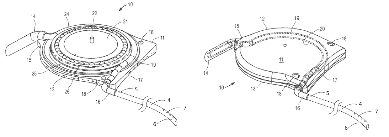
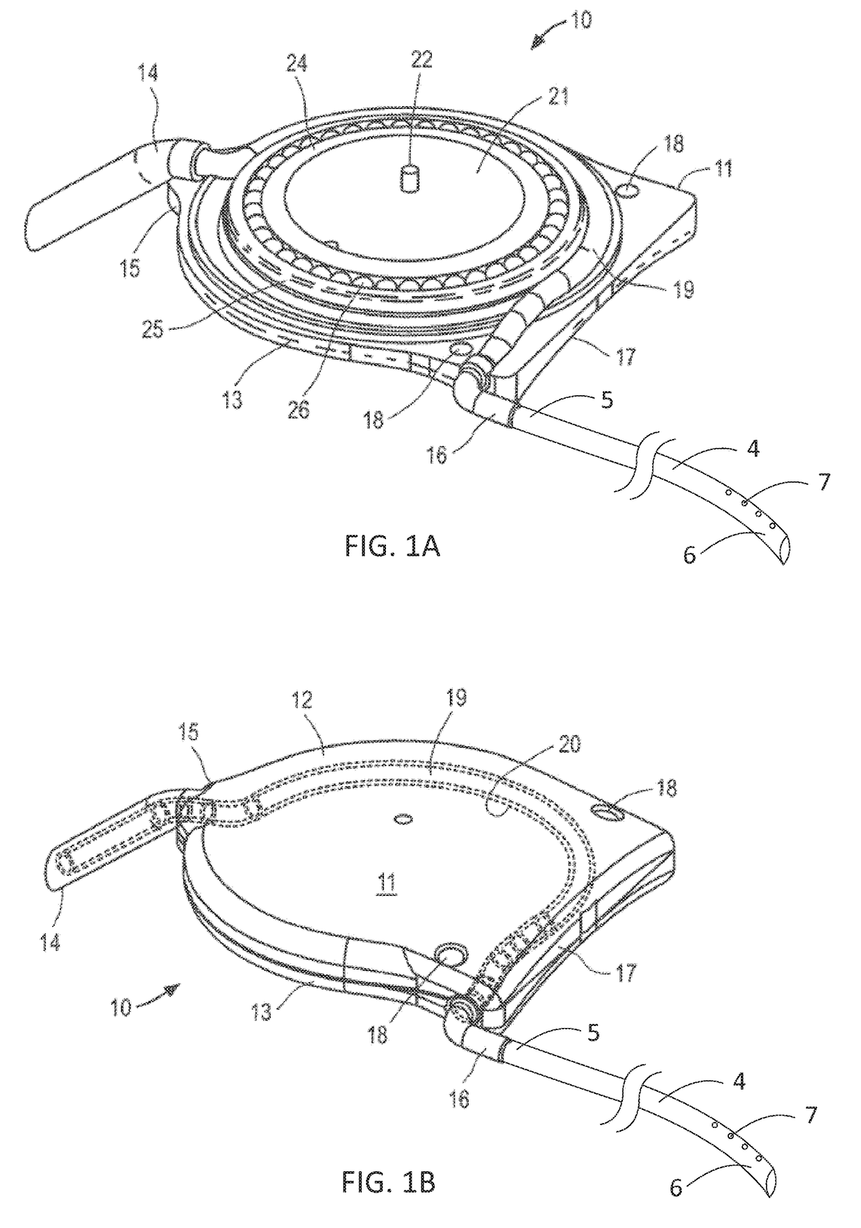
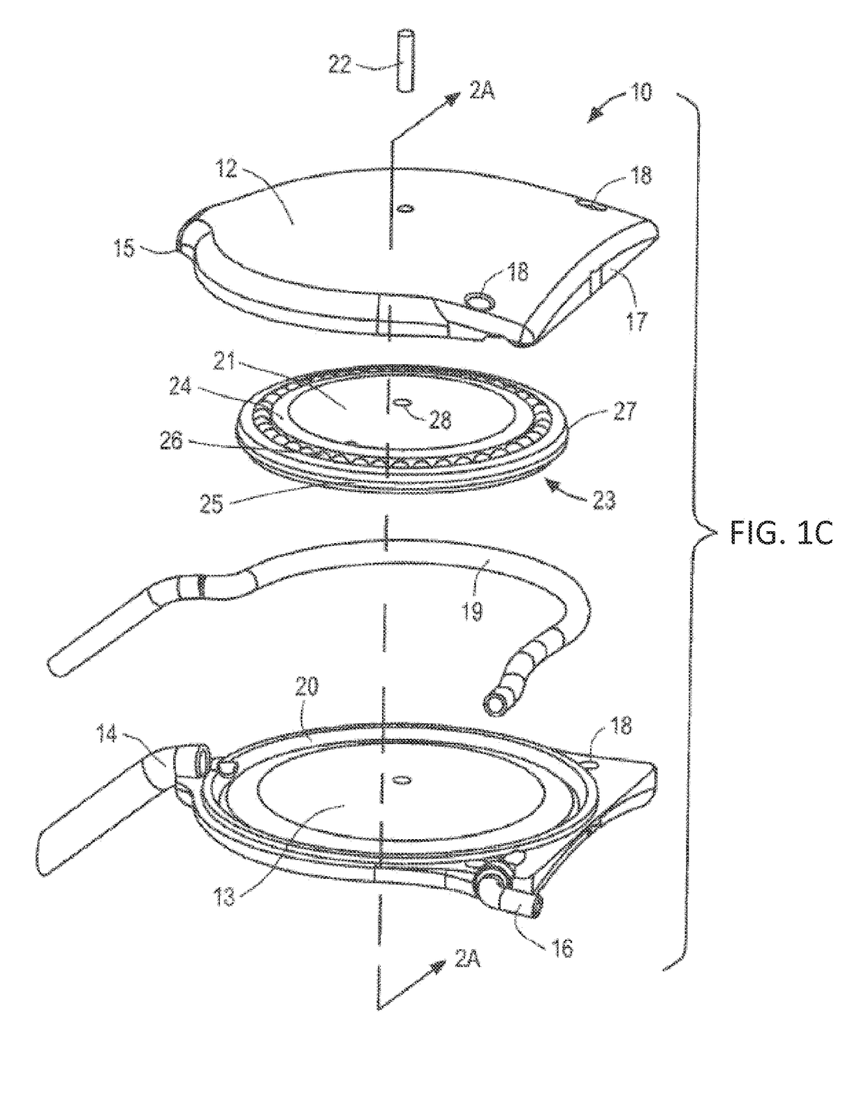
[0037]The ocular drainage system of the present invention comprises an implantable device having a valve that may be non-invasively adjusted to control the resistance to flow of aqueous humor from the anterior chamber of the eye, through the valve, and to a sink outside the eye (e.g., a bleb formed under a scleral flap or the orbital fat space of the eye). For example, as shown in FIGS. 1A and 1B, the implantable device is coupled to a flexible drainage tube having a distal end disposed within an orbital fat space of the eye and a proximal end coupled to an outlet port of the implantable device, so that aqueous humor flows from the anterior chamber of the eye, through the valve and flexible drainage tube, and is deposited in the orbital fat space of the eye. The ocular drainage system further comprises an external control unit that permits a health care provider to periodically adjust the valve within the implantable device to maintain intraocular pressures within a desired range, t...


PUM


Abstract

An ocular drainage system is provided for treating diseases that produce elevated intraocular pressures, such as glaucoma, wherein the system includes an implantable device and an external control unit, the implantable device includes a non-invasively adjustable valve featuring at least one deformable tube and a disk rotatably mounted within a housing, such that rotation of the disk using the external control unit causes the disk to apply a selected amount of compression to the deformable tube, thereby adjusting the fluidic resistance of the deformable tube and regulating the intraocular pressure.

Description

CROSS-REFERENCE TO RELATED APPLICATIONS[0001]This application is a continuation-in-part application of U.S. application Ser. No. 14 / 819,286, filed Aug. 5, 2015, now U.S. Pat. No. 9,655,779, which is a continuation application of U.S. application Ser. No. 13 / 349,353, filed Jan. 12, 2012, now U.S. Pat. No. 9,101,445, which claims the benefit of priority of U.S. Provisional Application No. 61 / 433,131, filed Jan. 14, 2011, the entire contents of each of which are incorporated by reference.FIELD OF INVENTION[0002]This application relates to apparatus and methods for draining excess intraocular fluid, for example, resulting from glaucoma.BACKGROUND OF THE INVENTION[0003]Glaucoma affects about 70 million people worldwide, and is a disorder associated with high pressure in the eye resulting in the generation of excess intraocular fluid (aqueous humor). Aqueous humor is produced at a rate of 2-3 μl / min by the ciliary body and it maintains constant intraocular pressure (IOP) around 12-20 mm H...

Claims


Application Information

Patent Timeline
Patent Type & Authority Applications(United States)
IPC IPC(8): A61F9/007A61M1/10A61F9/00A61M27/00F04B43/04
CPCA61F9/00781A61M2210/0612A61M27/002A61F9/00736A61F9/0008F04B43/04A61F9/00A61M1/1037F04B43/09F04B43/1253F04B2201/1208F04B43/14
Inventor BIGLER, STEPHANESTERGIOPULOS, NIKOLAOS
Owner ECOLE POLYTECHNIQUE FEDERALE DE LAUSANNE (EPFL)
Why Eureka
Try Eureka