Computerized optical analysis methods of mr (magnetic resonance) images for quantifying or determining liver lesions

a technology of computerized optical analysis and magnetic resonance, applied in the field of hepatic diagnosis, can solve the problems of insufficient sensitiveness and often inaccurate, and the performance of this kind of test may be insufficient, so as to monitor the potential therapeutic

Inactive Publication Date: 2018-02-22
SERVICIO ANDALUZ DE SALUD (SAS) +2
View PDF0 Cites 2 Cited by
  • Summary
  • Abstract
  • Description
  • Claims
  • Application Information

AI Technical Summary

Benefits of technology

[0008]The present invention provides computerized optical analysis methods of conventional non-contrast-enhanced MR (magnetic resonance) images of the liver, said methods enable the detection of steatohepatitis by NASH-MRI and significant fibrosis by Fibro-MRI in patients suffering from NAFLD. By using the methods of the present invention it is possible to forecast the rate of disease progression, to support therapeutic decision-making, and to monitor potential therapeutic effects.

Problems solved by technology

Although biopsies can provide important information regarding the degree of liver damage, in particular the severity of necro-inflammatory activity, fibrosis and steatosis, the procedure also presents several limitations, such as sampling error, invasiveness, cost, pain for patients which in turn brings forth a certain reluctance to undergo such a procedure; and finally complications may arise from such procedure, which in some cases can even lead to mortality.
As a result, it is not sensitive enough and often inaccurate in patients with advanced fibrosis.
Finally, it is generally admitted that around half of all FLD cases are detected by usual blood tests and around half by ultrasonography, resulting in around one quarter of missed diagnosis when both are used.
However, according to the French National Agency for Health (HAS), updated in December 2008, and current international opinion, the performance of this kind of test may be insufficient, especially due to the reference based on a subjective grading of liver steatosis with a poor inter-observer reproducibility.

Method used


Image

  • Computerized optical analysis methods of mr (magnetic resonance) images for quantifying or determining liver lesions
  • Computerized optical analysis methods of mr (magnetic resonance) images for quantifying or determining liver lesions
  • Computerized optical analysis methods of mr (magnetic resonance) images for quantifying or determining liver lesions

Examples


example 1

nt and Standardization of NASH-MRI for Detection of Steatohepatitis

[0154]Estimator E3 (harmonic mean) from MRI protocol SSFSE-T2, estimator E57 (2-2 order contrast) from MRI protocol DYNAMIC, and estimator E73 (weighted mean curvature) from MRI protocol FAST-STIR, were found to be independently associated with NASH.

[0155]Model coefficients associated with each one of these independent variables were β1=0.079 (OR: 1.08, 95% CI: 1.02-1.15; p=0.015) and β2=0.22 (OR: 1.14, 95% CI: 1.03-1.26; p=0.015).

[0156]These estimators influence on the predictive equation to obtain the probability of suffering steatohepatitis:

NASH-MRI=1 / (1e(1.654−0.079*E3_T2−0.127*E57_DYN*E73_FAST))

[0157]AUROC obtained was 0.88 (95% CI: 0.77-0.99) in the estimation cohort. Values for sensitivity of 87%, specificity of 74%, positive predictive value (PPV) of 80% and negative predictive value (NPV) of 82% were obtained from prediction based on a cut-off point of 0.5 for NASHMRI.

[0158]In the validation cohort, NASH-MRI...

example 2

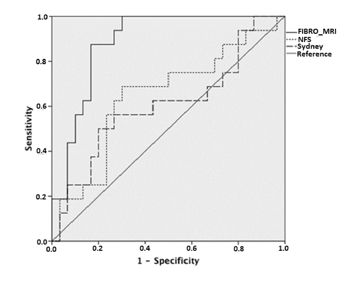
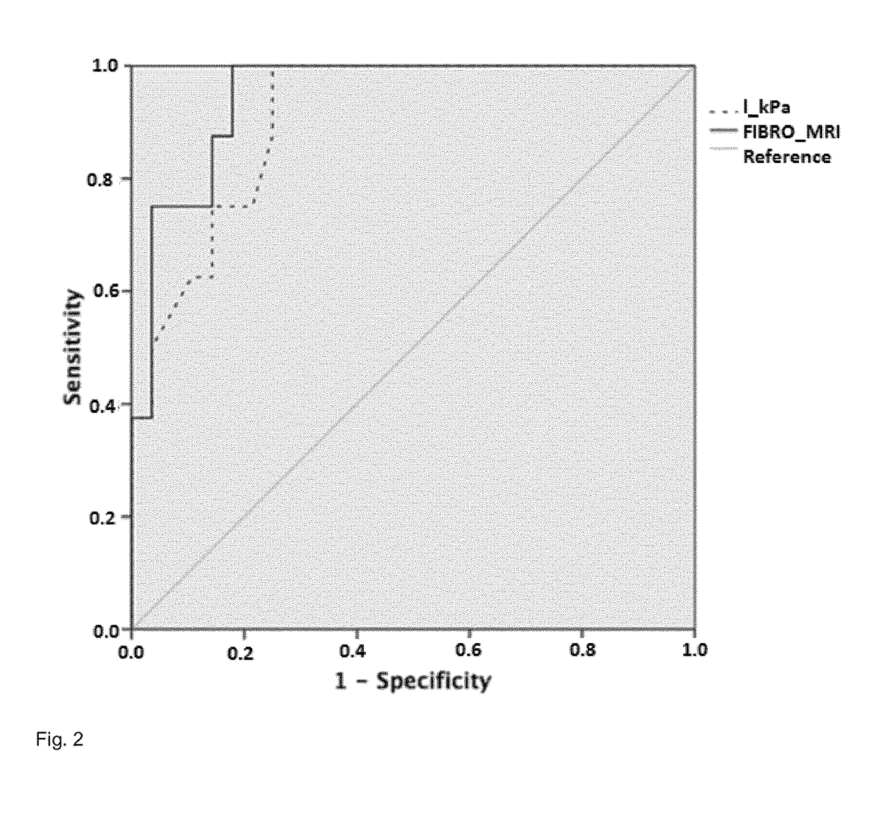
nt and Standardization of Fibro-MRI for Detection of Fibrosis

[0159]Estimator E22 (Pearson's asymmetry coefficient) from MRI protocol SSFSE-T2 and estimators E3 (harmonic mean), E6 (mode), E31 (column's mean of multi-oriented co-occurrence matrix) and E75 (maximum of main curvatures) from MRI protocol DYNAMIC were found to be independently associated with fibrosis. Model coefficients associated with each of these independent variables were: β1=1.101 (OR: 3.01, 95% CI: 1.25-7.25; p=0.014); β2=−1.105 (OR: 0.33, 95% CI: 0.14-0.77; p=0.010); β3=−115.737 (OR: 0.08, 95% CI: 0.02-0.14; p=0.046) β4=0.696 (OR: 2.00, 95% CI: 1.19-3.38; p=0.009); and β5=−0.825 (OR: 0.44,v95CI %: 0.21-0.93; p=0.030) should be introduced into the predictive equation to obtain the risk of suffering fibrosis as:

Fibro-MRI=1 / (1+e(−4.207-1.101*E3_DYN+1.105*E6_DYN+115.737*E22_T2−0.696*E31_DYN+0.825*E75_DYN))

[0160]In the estimation cohort, AUROC was 0.94 (95% CI: 0.87-1.00) with a sensitivity of 81%, specificity of 85%,...

example 3

zation of NASH-MRI and Fibro-MRI Across MRI Systems

[0162]NASHMRI, calculated using a GE scanner, showed a similar diagnostic accuracy AUROC=0.76 (95% CI: 0.58-0.96) vs. AUROC=0.83 (95% CI: 0.70-0.96); p=ns in comparison with NASHMRI calculated in patients that underwent MRI with the Philips system. With respect to FibroMRI, evaluations performed using a GE scanner showed an AUROC=0.81 (95% CI: 0.66-0.95) vs. AUROC=0.86 (95% CI: 0.72-0.99) using the Philips system (p=ns).

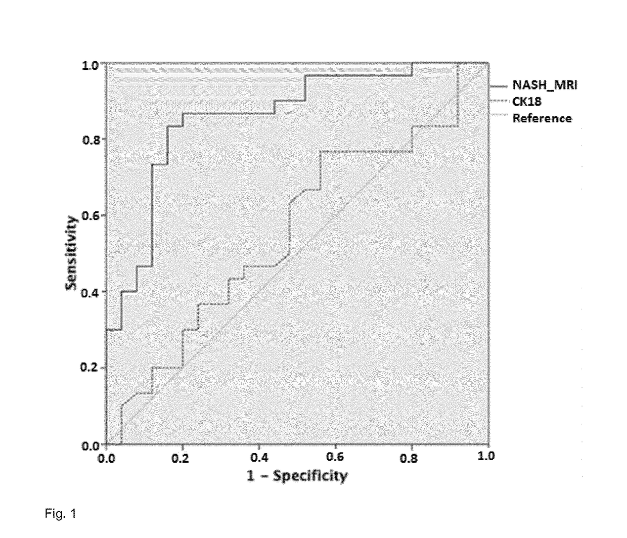
[0163]Comparative analysis with non-invasive biochemical markers of steatohepatitis NASHMRI was compared with FGF-21 and CK-18 in the diagnosis of steatohepatitis in a cohort of 64 patients. NASHMRI offered the best diagnostic accuracy with an AUROC of 0.86 (95% CI: 0.76-0.96) for steatohepatitis presence significantly better than CK-18 levels AUROC of 0.56 (95% CI: 0.40-0.71; p<0.05) (FIG. 1). NAS score correlated with both NASH-MRI (r=0.38; p<0.001) and CK-18 levels (r=0.29; p<0.02).

[0164]Comparative analysis with ...


PUM


Abstract

A method computerized optically analyzes methods of conventional, preferably non-contrast-enhanced, MR (magnetic resonance) images of the liver. The method enables the detection of steatohepatitis by NASH-MRI and significant fibrosis by Fibro-MRI in patients suffering from NAFLD. The method enables forecasting the rate of disease progression, to support therapeutic decision-making, and to monitor potential therapeutic effects.

Description

FIELD OF THE INVENTION[0001]This invention relates to the field of hepatic diagnosis and more precisely to computerized optical analysis methods of conventional, preferably non-contrast-enhanced, MR (magnetic resonance) images for quantifying or determining liver lesions, especially due or related to liver impairment, liver steatosis, non-alcoholic fatty liver disease (NAFLD), or non-alcoholic steatohepatitis (NASH).BACKGROUND OF THE INVENTION[0002]FLD (Fatty Liver Disease) describes a wide range of potentially reversible conditions involving the liver, wherein large vacuoles of triglyceride fat accumulate in hepatocytes via the process of steatosis (i.e. the abnormal retention of lipids within a cell). FLD is commonly associated with alcohol or metabolic syndromes (diabetes, hypertension, dyslipidemia, abetalipoproteinemia, glycogen storage diseases, Weber-Christian disease, Wolman disease, acute fatty liver of pregnancy, lipodystrophy). However, it can also be due to nutritional c...

Claims


Application Information

Patent Timeline
Patent Type & Authority Applications(United States)
IPC IPC(8): G01R33/56A61B5/055A61B5/00G06T7/00G01R33/561
CPCG01R33/5608A61B5/055A61B5/4244G06T7/0012G01R33/5602G01R33/5607G01R33/5601G01R33/5617G06T2207/10088G06T2207/30056
Inventor ROMERO GOMEZ, MANUELGOMEZ GONZALEZ, EMILIOGALLEGO DURAN, ROCIOCERRO SALIDO, PABLO
Owner SERVICIO ANDALUZ DE SALUD (SAS)
Why Eureka
Try Eureka