Methods and systems for computed tomography

a computed tomography and computed tomography technology, applied in tomography, diagnostic recording/measuring, applications, etc., can solve problems such as reducing the accuracy of perfusion analysis

Active Publication Date: 2018-08-30
GENERAL ELECTRIC CO
View PDF3 Cites 1 Cited by
  • Summary
  • Abstract
  • Description
  • Claims
  • Application Information

AI Technical Summary

Benefits of technology

The patent text describes a method for reconstructing an image from projection data obtained during a scan. The method is based on a model that relates the timing of an event to be imaged to the person's heart rate during the scan. By using this model, the timing of the reconstruction can be consistent for a series of reconstructions, resulting in a more reliable and accurate registration of the images.

Problems solved by technology

Displacements in the anatomy from image to image or volume to volume will introduce errors and reduce the accuracy of the perfusion analysis.

Method used


Image

  • Methods and systems for computed tomography
  • Methods and systems for computed tomography
  • Methods and systems for computed tomography

Examples


Embodiment Construction

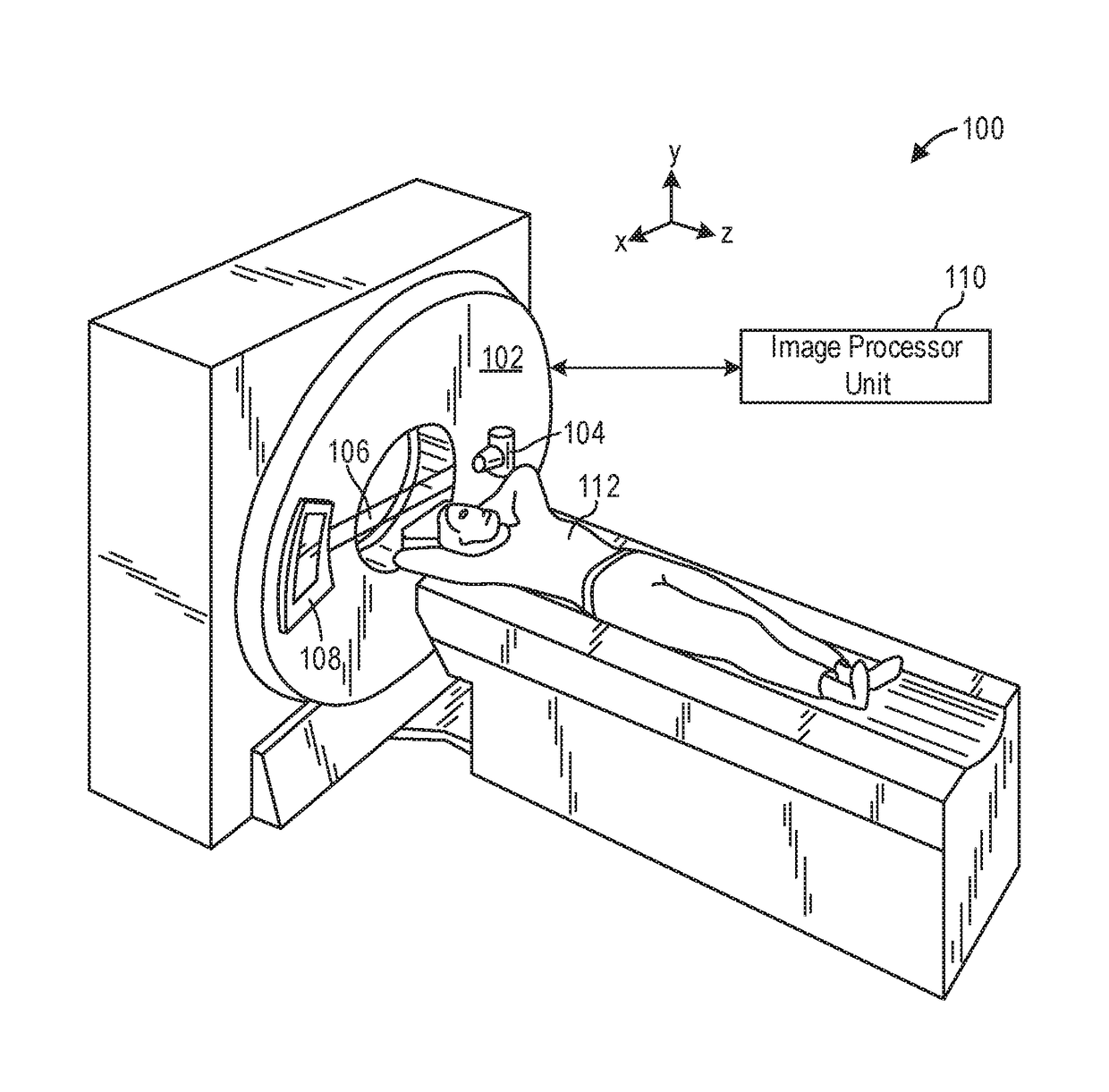
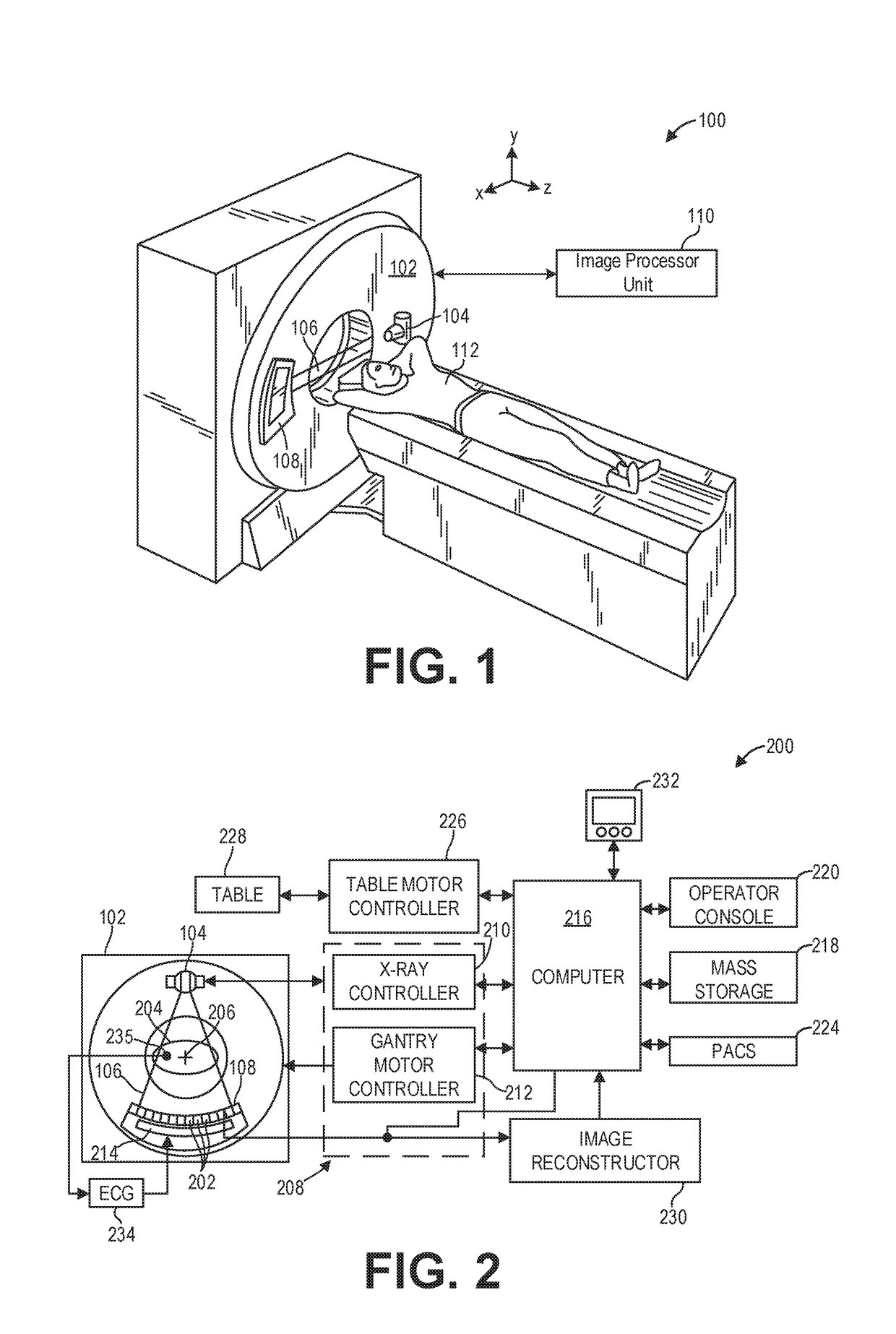
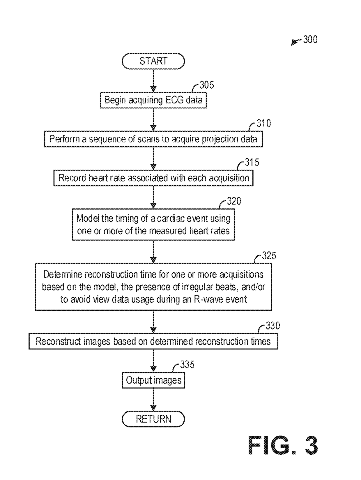
[0012]The following description relates to various embodiments of medical imaging system. In particular, methods and systems are provided for imaging an organ such as a heart with an imaging system, such as the CT imaging system depicted in FIGS. 1 and 2. A method for consistently imaging an organ, such as the method depicted in FIG. 3 and illustrated in FIG. 4, may include dynamically determining a reconstruction time for a series of reconstructions so that the reconstructions are best registered. Additionally, a method for obtaining consistent image reconstructions in the presence of irregular heartbeats, such as the method depicted in FIG. 5, may include reconstructing multiple images from data acquired during the irregular cardiac cycle, and selecting the image that best registers with neighboring frames.

[0013]Though a CT system is described by way of example, it should be understood that the present techniques may also be useful when applied to images acquired using other imagi...


PUM


Abstract

Methods and systems are provided for cardiac computed tomography imaging. In one embodiment, a method comprises reconstructing an image from projection data acquired during a scan with a reconstruction time determined based on a model relating a timing of an event to be imaged to a heart rate measured during the scan. In this way, the timing of a reconstruction may be consistently applied for a series of reconstructions, thereby inherently registering the reconstructions.

Description

FIELD[0001]Embodiments of the subject matter disclosed herein relate to non-invasive diagnostic imaging, and more particularly, to image reconstruction for cardiac computed tomography (CT) imaging systems.BACKGROUND[0002]Non-invasive imaging technologies allow images of the internal structures of a patient or object to be obtained without performing an invasive procedure on the patient or object. In particular, technologies such as computed tomography (CT) use various physical principles, such as the differential transmission of x-rays through the target volume, to acquire image data and to construct tomographic images (e.g., three-dimensional representations of the interior of the human body or of other imaged structures).[0003]Dynamic perfusion of the heart, liver, or other organs requires that images be generated over a period of time as a contrast agent moves through the organ. Analysis of the variation in the localized contrast agent is then used to infer any of a number of per...

Claims


Application Information

Patent Timeline
IPC IPC(8): A61B6/00A61B5/0456A61B5/0245A61B6/03A61B5/04A61B5/352
CPCA61B6/5205A61B5/0456A61B5/0245A61B6/4435A61B5/04012A61B6/481A61B6/032A61B5/7292A61B6/486A61B6/503A61B6/507A61B6/5211A61B6/5288G16H50/20A61B5/349
Inventor JACKSON, JOHN IRVINWOODFORD, MARK EDWARD
Owner GENERAL ELECTRIC CO
Why Eureka
Try Eureka