Non-Invasive Breast Cancer Detection Using Co-Registered Multimodal Probes: Microwave Nearfield Radar Imaging (NRI), Digital Breast Tomosynthesis (DBT), Ultrasound Imaging (US) And Thermoacoustic Imaging (TA)

a multi-modal, breast cancer technology, applied in the field of non-invasive breast cancer detection using co-registered multi-modal probes, can solve the problems of increasing the risk of breast cancer, increasing the difficulty of detecting breast cancer, and different chances of detecting early breast cancer, so as to achieve high fibrous-to-cancerous contrast, high resolution, and high sensitivity and specificity.

Inactive Publication Date: 2018-10-04
NORTHEASTERN UNIV
View PDF0 Cites 3 Cited by
  • Summary
  • Abstract
  • Description
  • Claims
  • Application Information

AI Technical Summary

Benefits of technology

[0025]4. Using an NM / UST / TAT or the NRI / USI modalities avoids the use of ionizing radiation associated with X-ray based DBT, while keeping high resolution and high fibrous-to-cancerous contrast, and thus providing high sensitivity and specificity.

Problems solved by technology

Depending on the breast tissue composition, however, the chances of detecting early breast cancer are different when using conventional screening mammograms.
Having breasts with relatively dense tissue (i.e., level 3 and 4) not only may increase the risk of getting breast cancer, but may also increase the difficulty of detecting breast cancer when using two-dimensional (2D), x-ray based screening mammograms.
Nevertheless, the number of unnecessary biopsies resulting from ultrasound imaging increases to an unacceptable level when it is used within a general population (i.e., dense and not dense breast) as a screening test.
Another adjunctive imaging modality is the bilateral breast magnetic resonance imaging (MRI), although MRI requires an intravenous injection of a contrasting agent and introduces an elevated cost.
First, existing modalities are not optimal to analyze all four levels of breast density (L1-L4).
Second, they require the use of either ionizing radiation (e.g., X-ray based), which may have potential carcinogenic effects, or potentially hazardous contrasting agents (e.g., MRI-based).
Third, current CM requires the use of adjunctive imaging modalities for dense breasts (L3-L4), and adjunctive imaging may result in additional cost, unnecessary anxiety and biopsies.
Fourth, current imaging modalities and their associated data analytic tools do not take into consideration the particular specifics of the patients, such as positive genetic mutations (e.g., BRCA1 and BRCA2), breast density, family history, among others.
Fifth, current modalities are not capable of providing low cost and size equipment that features high contrast and resolution.
Sixth, in the case of multimodal systems that provide additional information concerning the breast tissue composition (such as density, compressibility, viscosity, dielectric constant, and conductivity) to 2D / 3D mammography or MRI, such multimodal systems are not capable of collecting all data in a single and quick session, and in a co-registered fashion.
Unfortunately, these systems both suffer from the aforementioned low radiological contrast between healthy breast tissue and cancerous tissue.
As a result, these technologies tend to produce a large number of false positives when used for early detection.
Unfortunately, standalone NRI systems typically struggle to accurately detect cancerous lesions due to the heterogeneous distribution of tissues within the breast, having a wide dynamic range in the relative dielectric constants that ranges from 5 (fatty) up to 55 (fibrous) for healthy tissues, and about 60 for cancerous tissues at around 1 GHz.

Method used


Image

  • Non-Invasive Breast Cancer Detection Using Co-Registered Multimodal Probes: Microwave Nearfield Radar Imaging (NRI), Digital Breast Tomosynthesis (DBT), Ultrasound Imaging (US) And Thermoacoustic Imaging (TA)
  • Non-Invasive Breast Cancer Detection Using Co-Registered Multimodal Probes: Microwave Nearfield Radar Imaging (NRI), Digital Breast Tomosynthesis (DBT), Ultrasound Imaging (US) And Thermoacoustic Imaging (TA)
  • Non-Invasive Breast Cancer Detection Using Co-Registered Multimodal Probes: Microwave Nearfield Radar Imaging (NRI), Digital Breast Tomosynthesis (DBT), Ultrasound Imaging (US) And Thermoacoustic Imaging (TA)

Examples


Embodiment Construction

[0056]A description of example embodiments of the invention follows.

[0057]The teachings of all patents, published applications and references cited herein are incorporated by reference in their entirety.

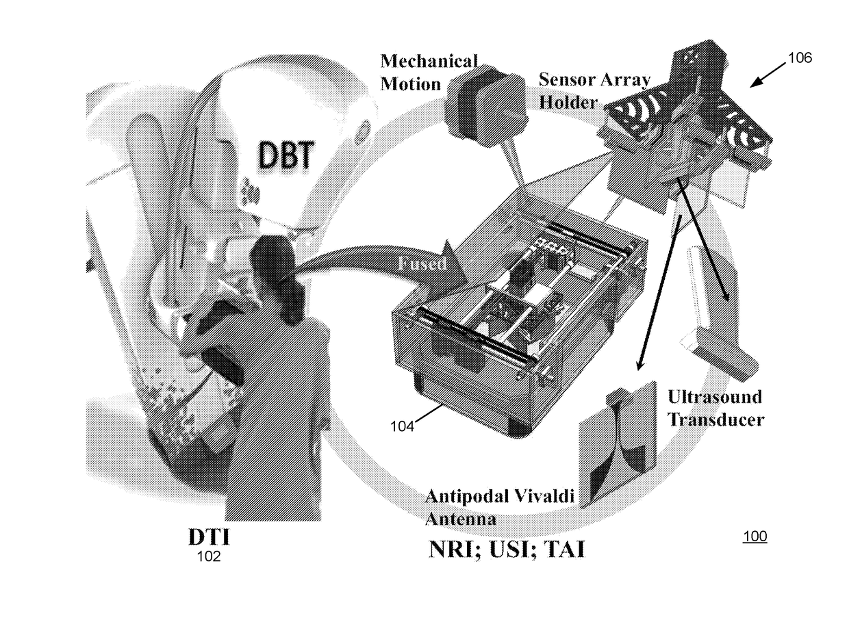
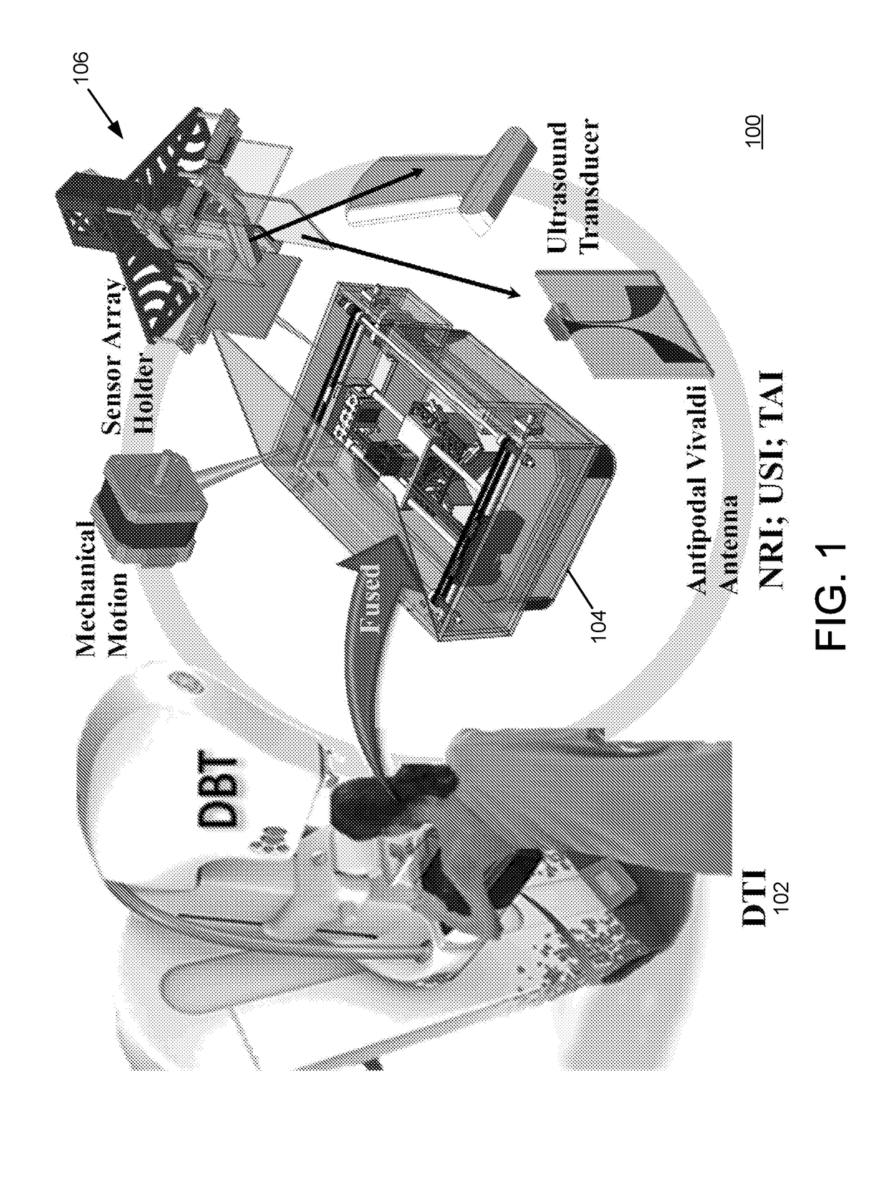
[0058]The described embodiments are directed to a breast cancer detection system that uses a multimodal imaging configuration. The described embodiments may utilize a fusion of two or more imaging modes, including for example (i) Digital Breast Tomosynthesis (DBT), (ii) Microwave Nearfield Radar Imaging (NRI), (iii) Ultrasound Imaging (USI) and Thermoacoustic Imaging (TAI). The described embodiments may evaluate the captured multimodal scan data jointly rather than independently. The described embodiments may further utilize co-registration of the two or more imaging modes, which ensures that the scans of all modes are captured with respect to the same physical configuration of the breast under study, i.e., while the breast is under clinical compression. The co-registration avoids th...


PUM


Abstract

A cancer detection system may comprise at least two imaging systems, each of which implements a different imaging modality, and each of which provides sampled image data. The system may further include, for each imaging modality, a modeling unit to produce modeled image data based on a common set of biophysical parameters. The system may also include a joint non-linear inversion module to receive information from each modeling unit and reconstruct a set of joint biophysical properties. The system may include a scaling unit to revise the common set of biophysical parameters based on the set of joint biophysical properties. The system may include a comparator to compare the sampled image data from each of the imaging systems to the corresponding modeled image data to determine a difference between the sampled image data and the modeled image data, and to determine when the difference is less than a threshold difference.

Description

RELATED APPLICATION[0001]This application claims the benefit of and priority to U.S. Provisional Application No. 62 / 413,873, filed on Oct. 27, 2016, U.S. Provisional Application No. 62 / 412,671, filed on Oct. 25, 2016 and U.S. Provisional Application No. 62 / 248,041, filed on Oct. 29, 2015. The entire teachings of the above applications are incorporated herein by reference.BACKGROUND[0002]Conventional mammography screening is the only modality that has been shown to reduce the chance of death from breast cancer in randomized control trials. Depending on the breast tissue composition, however, the chances of detecting early breast cancer are different when using conventional screening mammograms.[0003]The breast is made up of a mixture of tissues, including fibrous connective and glandular tissues, as well as fatty tissue. Radiologists classify breast density using a four level density scale, L1 through L4, where L1 describes the lowest breast density and L4 describes the highest breas...

Claims


Application Information

Patent Timeline
Patent Type & Authority Applications(United States)
IPC IPC(8): A61B6/00G06T7/00G06T11/00A61B5/05A61B6/02A61B8/08A61B5/00
CPCA61B6/5247G06T7/0014G06T11/008A61B5/0507A61B6/025A61B6/502A61B6/5217A61B8/0825A61B5/0093A61B5/7267G06T2207/10084G06T2207/20081G06T2207/30068G06T2207/10072G06T2207/10116G06T2207/10132
Inventor MARTINEZ-LORENZO, JOSE A.OBERMEIER, RICHARDGHANBARZADEH, ASHKANMOLAEI, ALIJUESAS, JUAN HEREDIA
Owner NORTHEASTERN UNIV
Why Eureka
Try Eureka