Device and method for medical imaging of coronary vessels

a technology of medical imaging and coronary arteries, applied in the field of devices and methods for medical imaging of coronary arteries, can solve problems such as important limitations, and achieve the effect of improving digital image processing

Active Publication Date: 2019-02-14
KONINKLJIJKE PHILIPS NV
View PDF0 Cites 6 Cited by
  • Summary
  • Abstract
  • Description
  • Claims
  • Application Information

AI Technical Summary

Benefits of technology

[0016]The present invention provides an approach to rely on a C-arm CT reconstruction of three-dimensional rotational angiography of the coronary arteries to generate a complete multiphase map. Indeed, accurate three-dimensional maps of the injected coronaries (over which the occluded segments are invisible) can be extracted from the three-dimensional rotational angiography, 3DRA, at any given heart phase.
[0016]The present invention provides an approach to rely on a C-arm CT reconstruction of three-dimensional rotational angiography of the coronary arteries to generate a complete multiphase map. Indeed, accurate three-dimensional maps of the injected coronaries (over which the occluded segments are invisible) can be extracted from the three-dimensional rotational angiography, 3DRA, at any given heart phase.
[0018]The present invention advantageously provides to rely on a set of second coronary vessel maps, which presents the opposite characteristics: they can be extracted accurately at any given heart phase, but they show only the vessel parts that are injected—and thus miss the occluded parts. These second coronary maps can be gained from a 3DRA, typically an X-per swing acquisition or any other single or dual axis rotational coronary angiography.
[0029]According to an exemplary embodiment of the invention, at the reference cardiac phase, the step of registering from the first map to the set of second maps is conducted. This advantageously provides to create coronary maps that combine the advantage of the CTA-based and the 3DRA-based maps.
[0020]The present invention advantageously protects the alignment of the CTA-based coronary map with the 3DRA-based coronary map; the coronary vessel maps could be aligned directly, without going through the two phase's motion compensation.

Problems solved by technology

The recanalization of chronically total occluded, CTO, coronary arteries is one of the most difficult percutaneous interventions, because the course of the occluded part of the vessel is invisible in angiography.
However, cardiac CTA are only performed at diastole in practice.
This is an important limitation for some applications like three-dimensional cardiac road-mapping in the case of CTO.

Method used


Image

  • Device and method for medical imaging of coronary vessels
  • Device and method for medical imaging of coronary vessels
  • Device and method for medical imaging of coronary vessels

Examples


Embodiment Construction

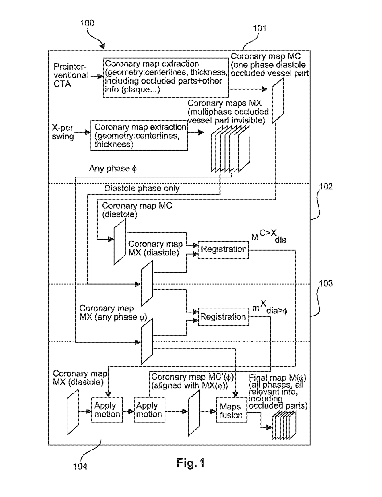
[0043]The illustration in the drawings is schematically and not to scale. In different drawings, similar or identical elements are provided with the same reference numerals. Generally, identical parts, units, entities or steps are provided with the same reference symbols in the figures.

[0044]FIG. 1 shows a schematic flowchart diagram of a method for medical imaging of coronary vessels according to an exemplary embodiment of the invention.

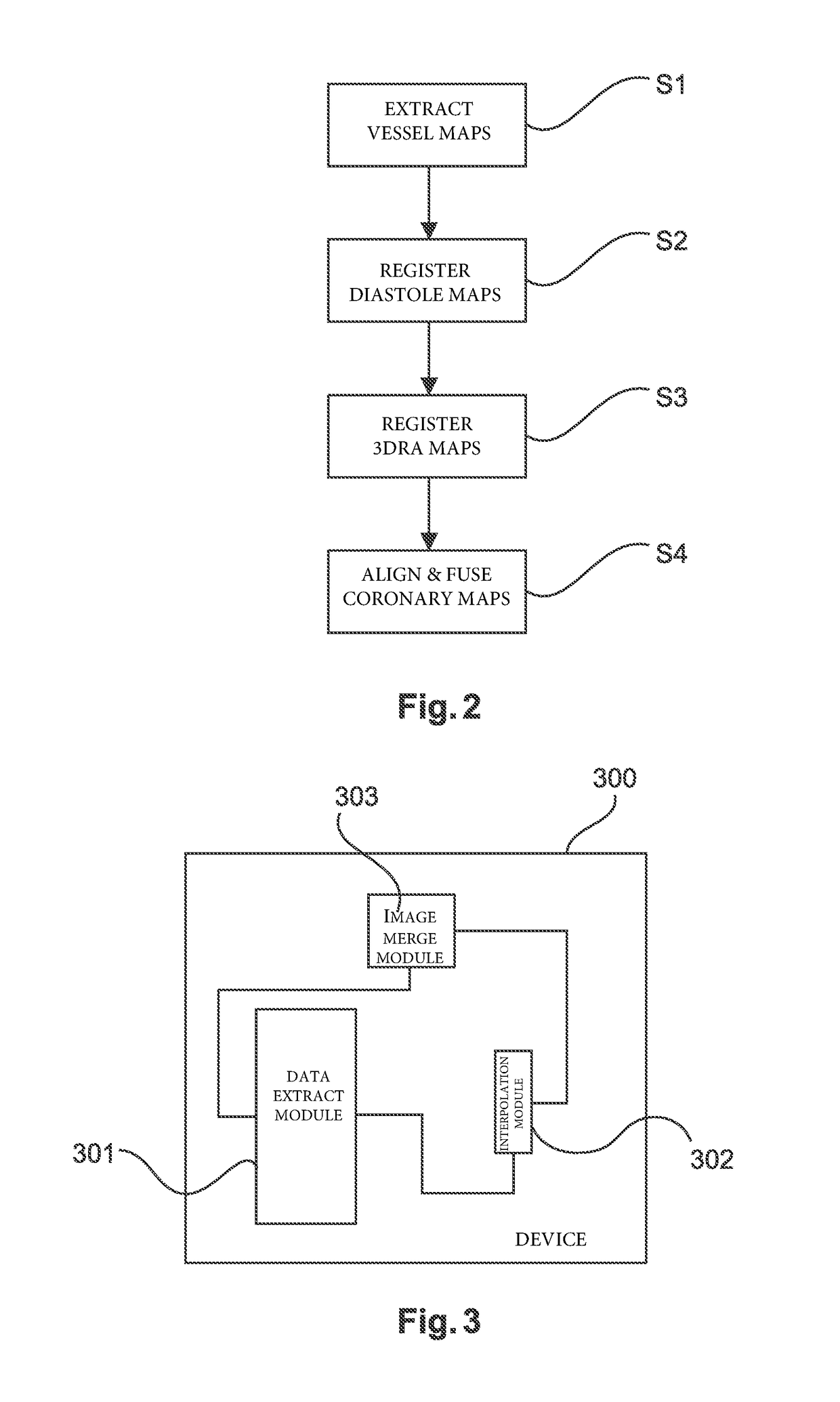
[0045]The method is visualized in terms of a function block diagram. A function block contains input variables, output variables, through variables, internal variables, and an internal behavior description of the function block. Function blocks are used primarily to specify the properties of a user function. Many software languages are based on function blocks.

[0046]The method or function block 100 may comprise, as sub-elements, four steps S1, S2, S3, S4 or four function blocks 101, 102, 103, 104:

[0047]In a first function block 101, corresponding to...


PUM


Abstract

The present invention relates to a method and a device for medical imaging of coronary vessels, the device comprising: a data extracting module configured to extract a first vessel map from computed tomography angiography data covering at least one reference cardiac phase and a set of second vessel maps from three-dimensional rotational angiography data covering at least one cardiac cycle; an interpolation module configured to generate a series of warped versions of the first vessel map aligned with the set of second vessel maps, the series starting at the at least one reference cardiac phase; and a merging module configured to merge the series and the set of second vessel maps at the different phases in order to generate a final imaging map of the coronary vessels.

Description

CROSS-REFERENCE TO PRIOR APPLICATIONS[0001]This application is a Continuation of application Ser. No. 15 / 127,972, filed Sep. 21, 2016, which is the U.S. National Phase application under 35 U.S.C. § 371 of International Application No. PCT / EP2015 / 055387, filed on Mar. 16, 2015, which claims the benefit of European Patent Application No. 14305429.4, filed on Mar. 26, 2014. These applications are hereby incorporated by reference herein.FIELD OF THE INVENTION[0002]The present invention relates to a device and a method for medical imaging of coronary vessels.BACKGROUND OF THE INVENTION[0003]U.S. Pat. No. 8,055,044 B2 describes a system for visualization of blood vessels and bones. One of two data sets, originating from two different radiological methods is processed to generate interim results, yielded by an operation on one of the data sets. The interim results are used to modify the other data set. Different imaging capabilities of the employed radiological methods promote a particular...

Claims


Application Information

Patent Timeline
Patent Type & Authority Applications(United States)
IPC IPC(8): G06T7/38A61B6/00
CPCA61B6/504G06T7/38A61B6/487A61B6/4441A61B6/5235A61B6/5288A61B6/032G06T2207/30101G06T2207/30048G06T2207/10081G06T7/33A61B6/503G06T2207/10076A61B5/0044A61B6/5247A61B5/0035G06T2207/10072A61B6/488
Inventor AUVRAY, VINCENT MAURICE ANDREFLORENT, RAOULLELONG, PIERRE HENRI
Owner KONINKLJIJKE PHILIPS NV
Why Eureka
Try Eureka