Flexible 3D rotational angiography and computed tomography fusion

a 3d rotational angiography and computed tomography technology, applied in the field of imaging systems, can solve the problems of overlapping property distributions of different types, affecting the segmentation of artery/vessel information in the 3d volume, and no absolute correct density value is available, so as to achieve the effect of sacrifying easy visual interpretation and visualizing ample volumes

Active Publication Date: 2011-11-08
KONINKLIJKE PHILIPS ELECTRONICS NV
View PDF14 Cites 12 Cited by
  • Summary
  • Abstract
  • Description
  • Claims
  • Application Information

AI Technical Summary

Benefits of technology

According to a preferred embodiment of the application, the system comprises means for applying a threshold to and filtering at least one of said data sets situated upstream of said means for registering. The means for registration can yield erroneous results, if the incoming data contains errors. Those errors are usually caused by artefacts originating in the acquisition process. An example of artefacts produced during the acquisition process are voxels that are filled with air, but are assigned a value that indicates a solid matter. If the other acquisition method is less prone to produce such artefacts, or produces different artefacts than those produced by the first acquisition method, then a registration algorithm risks to fail, because it calculates the positions, scales and orientations of the target object in each of the two data sets under the assumption, that the prevailing number of voxels with a high absorption coefficient value are part of the target object. The effectiveness of the means for registration can be restored by a thresholding method. Indeed, when the voxel values outside the skull are set to zero by thresholding on a value between the erroneous air voxel values and the grey matter or brain tissue values, the means for registration deliver data essentially artefact free.
The method's second acquisition method preferably is a three-dimensional rotational angiographic method. Three-dimensional rotational angiography (3DRA) is effectively used for visualization of high contrast artery / vessel structures.
The method according to a preferred embodiment further comprises a step of obtaining a sectional view of the first, second and / or a combination of both three-dimensional image data sets with a preselected geometrical plane. In order for a human expert to analyze the acquired data, it has to be displayed. The interpretation of the displayed volume data is greatly facilitated, when a sectional view of the data is provided in a meaningful manner, i.e. uninteresting parts of the volume are cut away by the section along a preselected geometrical plane.
Preferably, the method further comprises the step of displaying the combined three-dimensional data set along a predefined path, in particular a blood vessel. Consequently, the method provides a way to visualize ample volumes without sacrifying easy visual interpretation by displaying a sequence of sectional views, each view centered around a different location, that is part of a predefined path. The path can for example correspond to a blood vessel, so that a human observer has the impression of traveling along this particular blood vessel. The vessel being visualized together with the volume in its vicinity provides an efficient way of representing large quantities of data for downstream expert evaluation.
Merging data originating from different acquisition methods for analysis and visualization, each acquisition method being particularly qualified for a distinct diagnostical task and / or type of tissue, has the advantage that all methods benefit from interim results determined by a method, that is particularly well suited for a particular, required operation.

Problems solved by technology

The problem of overlapping property distributions for different types of tissue is particularly present in three-dimensional rotational angiography.
In general, in 3DRA no absolute correct density values will be available due to insufficient calibration and reconstruction procedures.
In this method, the segmentation of artery / vessel information in the 3DRA volume is hindered by the fact that the artery / vessel densities are in the same range as bone material, due to beam hardening caused by the skull.
Furthermore, the high density data of the skull in the CT volume, which is typically used for orientation of the CT slices, prevents a clear view on the arterial structures, when both volumes are combined.
While Computer Tomography (CT) is known to provide high contrast between bony structures and soft tissue (e.g. skull as opposed to brain), the use of three-dimensional rotational angiography (3DRA) for extraction of 3-D vessel information is hindered by the fact, that the intensity distribution of high intensity bone overlaps with the intensity distribution of contrast filled vessels.
This effect is especially due to the phenomenon of beam-hardening caused by the skull, in combination with insufficient calibration and reconstruction procedures.
However, the method only interprets the inherently unsuitable data material provided by the three-dimensional rotational angiographic acquisition method in a different manner and does not revert to more suitable information sources.

Method used


Image

  • Flexible 3D rotational angiography and computed tomography fusion
  • Flexible 3D rotational angiography and computed tomography fusion
  • Flexible 3D rotational angiography and computed tomography fusion

Examples


Embodiment Construction

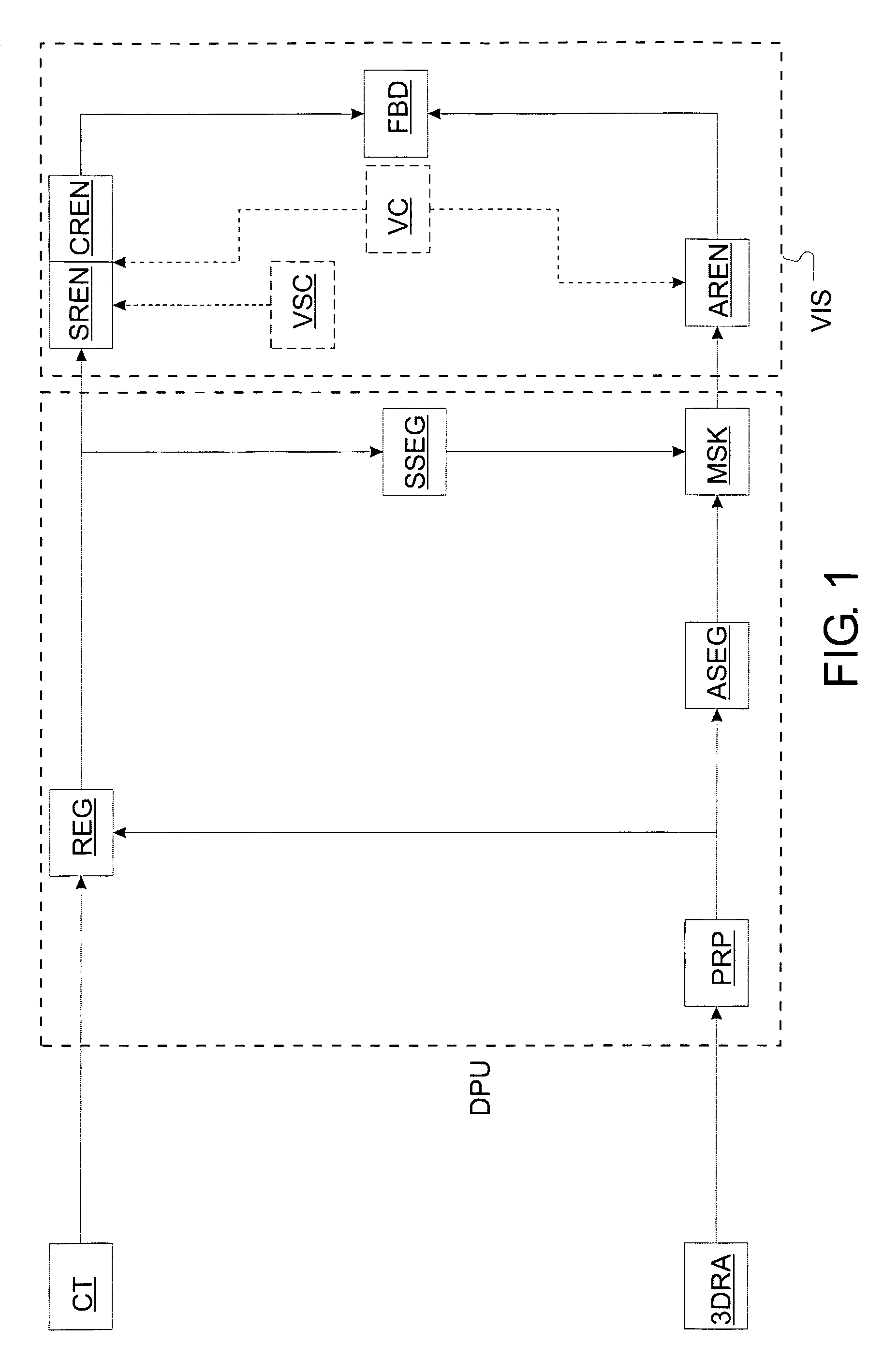
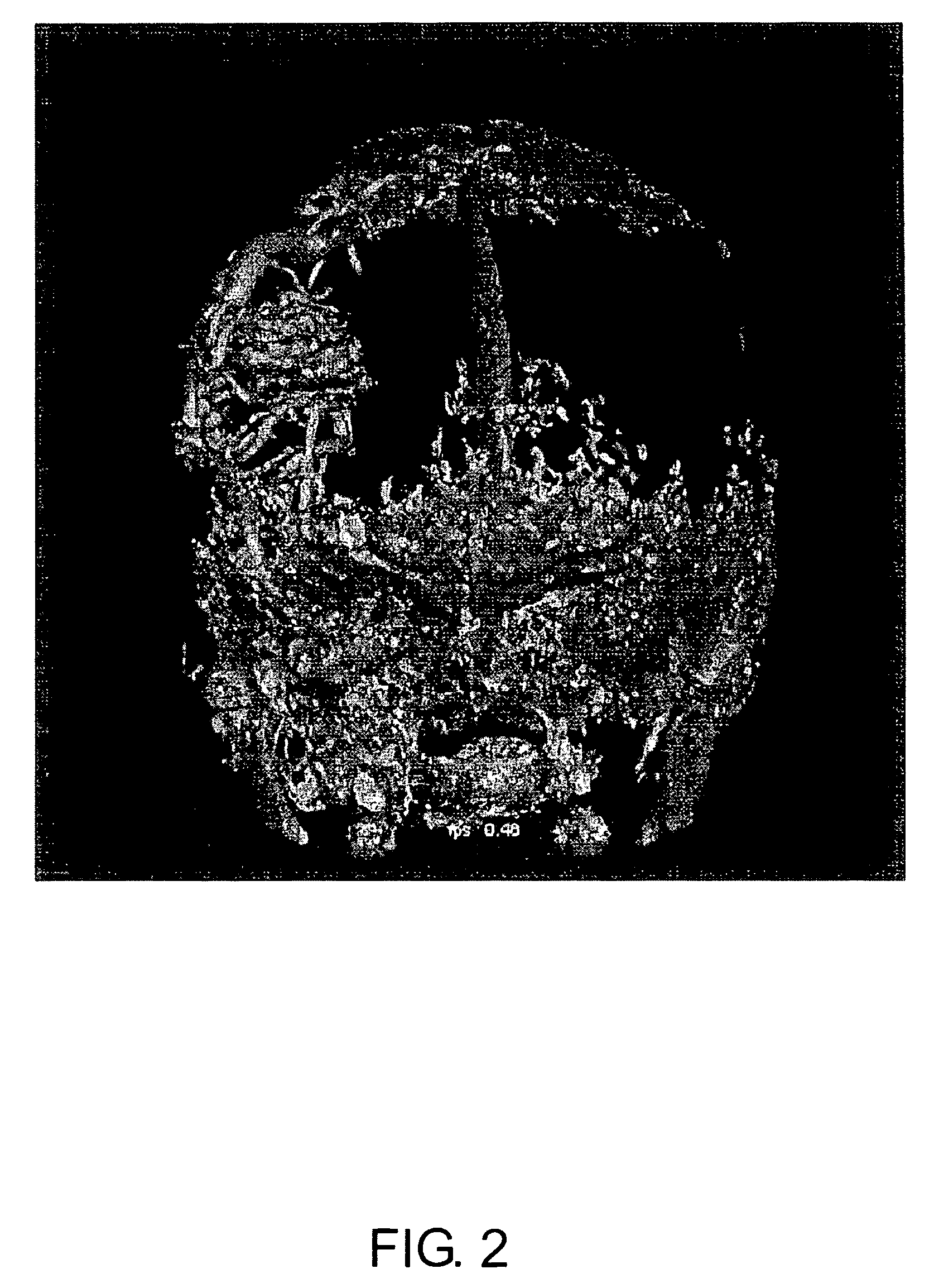
The present application is a system and method for flexible fusion of two data sets representing three-dimensional densities of a physical properties. According to a preferred embodiment of the present application, two data sets, which both represent the same object, but were generated using different acquisition methods, are combined in such a way, that the advantages of a particular acquisition method can be of use for a data set that was acquired using a different radiological method. For sake of clarity, the present application is explained here with reference to a preferred embodiment employing two diagnostical radiological methods in particular, these two methods being three-dimensional rotational angiography (3DRA) on the one hand and computer tomography (CT) on the other hand. CT is typically used for visualization of low contrast soft tissue such as brain material, while 3DRA is effectively used for visualization of high contrast artery / vessel structures. Since 3DRA and CT ...


PUM


Abstract

One of two data sets, originating from two different radiological methods is processed to generate interim results, yielded by an operation on one of the data sets. The interim results are used to modify the other data set. Different imaging capabilities of the employed radiological methods promote a particular task, e.g. the segmentation of a given type of tissue. When the combined data of both methods is displayed, clinical users benefit from the complementary information. Care is taken, that only relevant information is presented to the user, as to avoid irrelevant data obscuring any data of interest. Therefore, the data to be displayed is filtered based on content, e.g. the type of tissue, and on location. Three-dimensional computer tomography and three-dimensional rotational angiography are particularly applicable radiological methods.

Description

BACKGROUNDThe present application relates to imaging systems, particularly diagnostic imaging systems. It finds particular application in conjunction with an integrated three-dimensional rotational angiographic (3DRA) and computerized tomographic (CT) diagnostic imaging system and will be described with particular reference thereto. However, it should be appreciated, that the present application may also find application in conjunction with other types of multi-modality diagnostic imaging systems. The present application-is particularly useful for visualizing blood vessel structures around and inside the skull.For diagnostical purposes in a clinical environment, the separate visualization of the various represented tissue types when employing a radiological acquisition method can provide useful information about the clinical picture of a patient. With different radialogic methods being specialized on the visualization of one or a few kinds of tissues, the goal of displaying only use...

Claims


Application Information

Patent Timeline
Patent Type & Authority Patents(United States)
IPC IPC(8): G06K9/00
CPCA61B6/032A61B6/463A61B6/504A61B6/5235A61B6/466Y02E60/36
Inventor MIELEKAMP, PIETER MARIA
Owner KONINKLIJKE PHILIPS ELECTRONICS NV
Why Eureka
Try Eureka