
Affinity engineered serum protein carrier binding domain
- Summary
- Abstract
- Description
- Claims
- Application Information
AI Technical Summary
Benefits of technology
Problems solved by technology
Method used
Image

Examples
examples
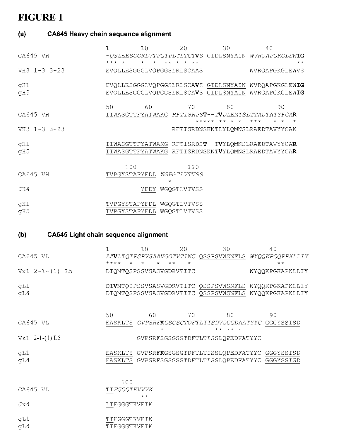
[0110]FIG. 1. Humanization and affinity reduction of antibody CA645. The heavy and light chain sequences of antibody CA645 are aligned with human germline acceptor framework sequences VH3 1-3 3-23 / JH4 and Vκ1 2-1-(1) L5 / Jκ4. Rabbit residues are in red, human residues are in black and CDRs are in blue (J-region CDR residues are shown but acceptor V-region CDRs are not). The grafted VH (gH) and VL (gL) sequences are shown below their corresponding human acceptor germline frameworks. Framework sequence differences between the rabbit and human framework sequences are shown with asterisks. Rabbit framework residues retained in the humanized grafts are highlighted in bold.
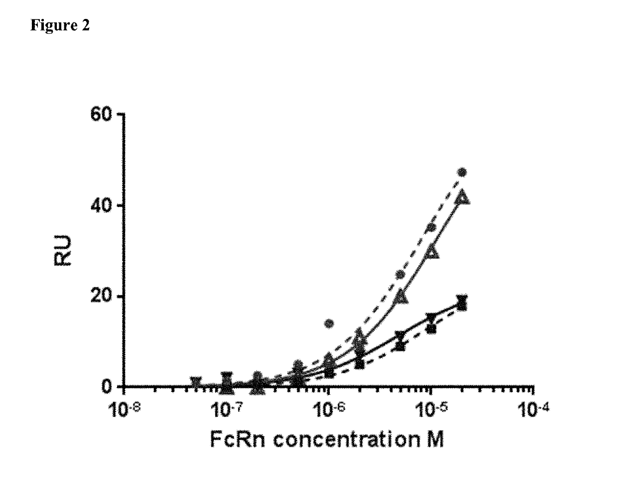
[0111]FIG. 2. Binding of FcRn to HSA and MSA in the presence or absence of CA645 gL4gH5 Fab. Binding to HSA in absence of Fab (red circle), binding to HSA in presence of Fab (red triangle), binding to MSA in absence of Fab (blue square), binding to MSA in presence of Fab (blue triangle).
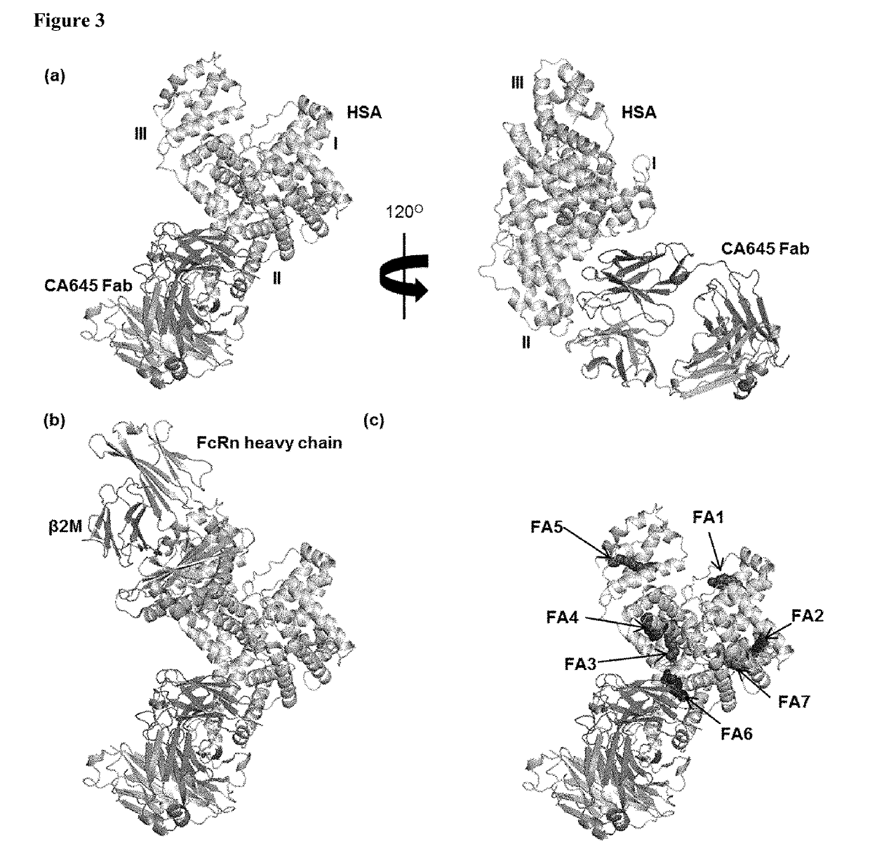
[0112]FIG. 3. (A) Crystal structur...
PUM
| Property | Measurement | Unit |
|---|---|---|
| Fraction | aaaaa | aaaaa |
| Fraction | aaaaa | aaaaa |
| Fraction | aaaaa | aaaaa |
Abstract
Description
Claims
Application Information