In situ visualization of kinase activity

a kinase activity and visualization technology, applied in the field of in situ visualization of kinase activity, can solve the problems of cellular and intracellular architecture disruption, limited detection of phosphorylated proteins, and requiring membrane permeabilization steps

Inactive Publication Date: 2019-06-20
DANA FARBER CANCER INST INC
View PDF0 Cites 0 Cited by
  • Summary
  • Abstract
  • Description
  • Claims
  • Application Information

AI Technical Summary

Benefits of technology

The patent describes methods for in situ detection of protein substrates of a specific kinase using an ATP analog. The methods involve incubating a sample with a fixative, an ATP analog, and a detection agent. The fixative can be an aldehyde such as formaldehyde or methanol. The ATP analog can be a thiophosphate or a fluorescent molecule. The methods can be used to visualize or quantify the activity of the kinase in a sample. The patent also provides kits for the methods.

Problems solved by technology

However, not all kinases have known substrates under a given condition, and detection of a phosphorylated protein is often limited by the availability of a potent and specific antibody.
However, the Shokat method requires a membrane permeabilization step due to the impermeability of bulky ATP analogs.
Upon membrane permeabilization, cellular and intracellular architectures are disrupted and the native biological context fails to be maintained.
Therefore, single-cell detection of kinase activity, which is commonly desired in state-of-the-art biological and biomedical research, has not been enabled in the original Shokat method.

Method used


Image

  • In situ visualization of kinase activity
  • In situ visualization of kinase activity
  • In situ visualization of kinase activity

Examples


example 1

of Substrates of a Kinase of Interest by Generating an Analog-Sensitive (AS) Mutation in the Kinase

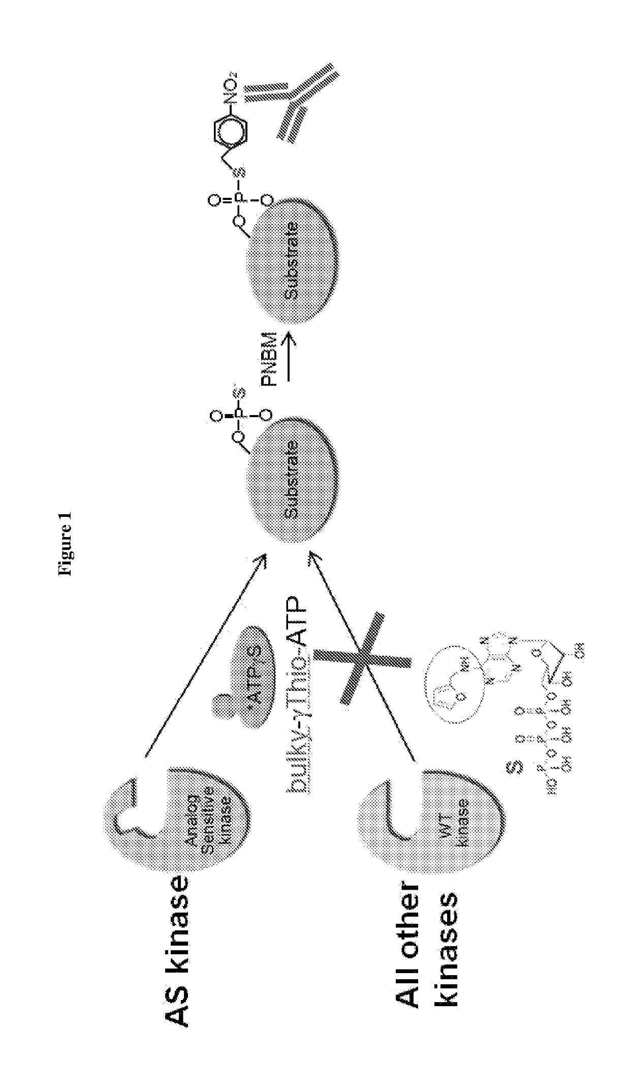
[0096]This example describes a method for detecting substrates of a kinase of interest. As shown in FIG. 1, the ATP-binding pocket of a kinase of interest was modified by a single amino acid substitution to generate an analog-sensitive (AS) enzyme which can accommodate a “bulky” analog of ATP. These analogs cannot be utilized by wild-type kinases. The bulky ATP was additionally modified to contain sulfur in the gamma phosphate, thereby being denoted “bulky-γ-Thio-ATP.” Cells or organisms e.g. mice were engineered to express the AS version of the kinase of interest. The AS kinase used bulky-γ-Thio-ATP and incorporated thiophosphate moieties into its protein substrates. These thiophosphate groups were next alkylated with p-Nitrobenzyl mesylate (PNBM) to create a semisynthetic epitope for an anti-thiophosphate ester antibody for detecting the substrates.

[0097]This method, modified from Sh...

example 2

of Cells in an In Vitro Culture for In Situ Visualization of Kinase Substrates

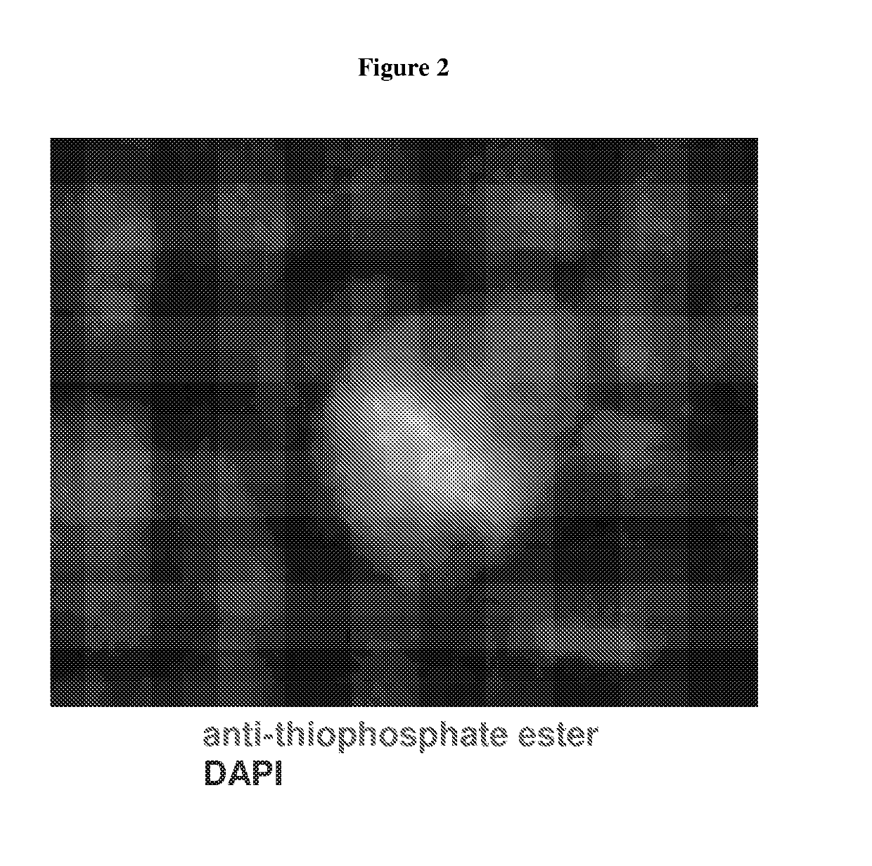
[0098]This example describes a method of fixing cells in an in vitro culture and visualizing substrates of an AS kinase in the cells, which resolved the problem in Example 1.

[0099]Murine CDK1 was engineered to comprise an AS mutation comprising M32V and F80G substitutions and this mutant CDK1 was substituted for the wild-type CDK1 in both alleles in murine embryonic stem cells. The sequence of the mutant CDK1 is shown in SEQ ID NO: 1, and other mutant CDK1 proteins comprising SEQ ID NOs: 2-4 are expected to function similarly Wild-type or AS mutant murine embryonic stem cells were cultured on glass coverslips and processed according to the steps in FIG. 5. The cells were fixed with 4% formaldehyde at room temperature for 5 minutes. The endogenous thiols were quenched with 20 mM iodoacetamide at 4° C. for 30 minutes. The cells were incubated with 100 μM furfuryladenosine-5′-O-(3-thiotriphosphate) in the pre...

example 3

of Brain Tissues for In Situ Visualization of Kinase Substrates

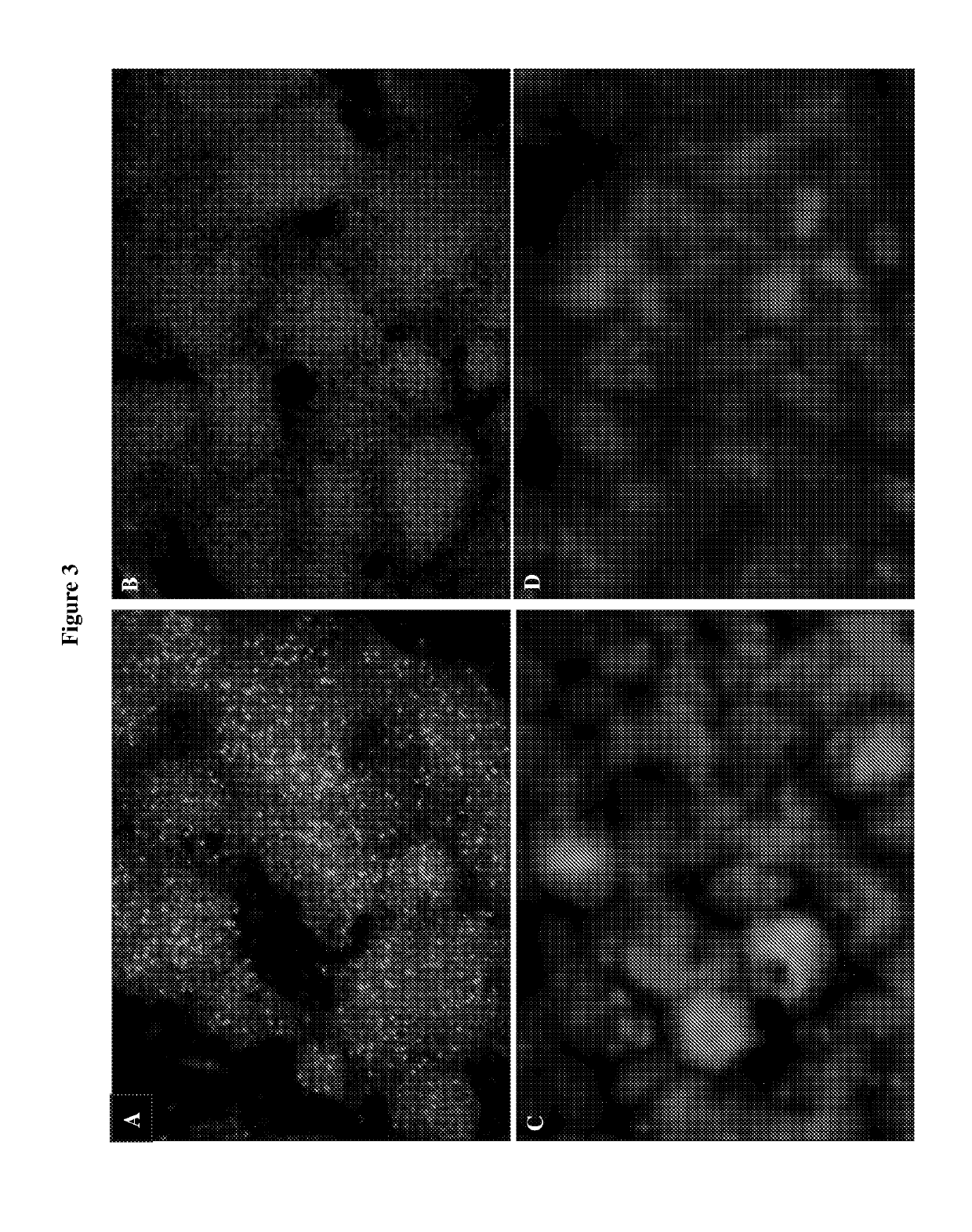
[0104]This example describes a method of fixing a tissue or an organ and visualizing substrates of an AS kinase in the tissue or organ, which resolved the problem in Example 1.

[0105]Murine CDK5 was engineered to comprise an AS mutation (F80G) and this mutant CDK5 was substituted for the wild-type CDK5 in both alleles in mice. The sequence of the mutant CDK5 is shown in SEQ ID NO: 9, and another mutant CDK1 protein comprising SEQ ID NO: 10 is expected to function similarly. Freshly harvested brains from adult wild-type or mutant mice were frozen and cut into 20 μm sections. The sections were mounted on glass coverslips and fixed with 4% formaldehyde for 5 minutes. The sections were then incubated with 100 μM furfuryladenosine-5′-O-(3-thiotriphosphate) in the presence of 0.1% Triton X-100 for 20 minutes, thereby allowing thiophosphorylation of substrates of the AS kinase. Thiophosphorylated substrates were alkylated with P...


PUM


Abstract

Kinases can be engineered to utilize an ATP analog that is not readily utilized by wild-type kinases by introducing a mutation in the ATP-binding pocket. However, application of this method has been limited by the membrane impermeability of the ATP analog. Provided herein are methods for in situ visualization of substrates of an analog-sensitive kinase, the method comprising a mild fixation step. Also provided herein are kits comprising a fixative, an ATP analog, and an agent for detecting the substrates modified by the ATP analog.

Description

RELATED APPLICATIONS[0001]This application claims the benefit of U.S. Provisional Application Nos. 62 / 377,262, filed Aug. 19, 2016, which is incorporated by reference herein in its entirety.GOVERNMENT SUPPORT[0002]This invention was made with government support under grants RO1 CA083688 and RO1 CA132740 awarded by the National Institutes of Health (NIH). The government has certain rights in the invention.BACKGROUND[0003]Phosphorylation is one of the most important protein modifications in signal transduction. Protein phosphorylation is regulated by protein kinases, which are under complex and dynamic regulations by other cellular apparatus. Methods have been developed to examine or monitor protein kinase activities, such as detecting phosphorylation of a known substrate protein. However, not all kinases have known substrates under a given condition, and detection of a phosphorylated protein is often limited by the availability of a potent and specific antibody. A more universal meth...

Claims


Application Information

Patent Timeline
IPC IPC(8): C12Q1/48
CPCC12Q1/485C12N9/12C12Y207/11C12Y207/11001C12Y207/11022C12Y207/11023G01N33/5088
Inventor SICINSKI, PETERMICHOWSKI, WOJCIECH
Owner DANA FARBER CANCER INST INC
Why Eureka
Try Eureka