
Device and method for enhanced visualization of the small intestine

Active Publication Date: 2019-07-25
NEPTUNE MEDICAL INC
View PDF0 Cites 2 Cited by
  • Summary
  • Abstract
  • Description
  • Claims
  • Application Information

AI Technical Summary

Benefits of technology

The patent describes a device for improving visualization in a body lumen during a medical examination. The device includes a capsule endoscope with a camera lens and a plurality of struts that extend from the capsule endoscope. The struts are designed to self-expand when the device is placed in the body lumen, creating a frame around the camera lens. This frame helps to improve the visibility of the body lumen during the examination. The device can be attached to the capsule endoscope using an annular ring and can include a tapered tip and a biodegradable portion. The technical effects of this device include improved visualization of the body lumen during a medical examination.

Problems solved by technology

Without such inflation, endoscopy moves through only a portion of an often-flattened lumen, failing to image significant parts of the lumen.
As a result, incomplete evaluation of the bowel is common when insufflation is not used.
However, traditional endoscopy is invasive, usually requires anesthesia, and can be very difficult to move through the small intestine.
However, none of these improvements are meaningful or helpful if the lumen is flattened and / or covers the lens.
For capsule endoscopy, luminal expansion techniques are currently not available.
b>. These exemplary scenarios can result in occlusion of the lens, a view of only the closed tissue, and / or an impartial view caused by blood, debris, or tissue interfering with the l
50%. Despite patient experience advantages relative to traditional endoscopic devices, these suboptimal rates have prevented the devices from reaching their pote
However, storing and methodically releasing pressurized air aboard a capsule in the gastrointestinal tract is problematic.
Excessive localized gas release can cause patient discomfort.
Chemical reactions struggle with heat, biocompatibility, foaming and bubbles, longevity, and adequate volume.
Capsule endoscopes including built-in radial extensions have been proposed as a means of making the device more lumen-centric to improve imaging, but these structures do not serve to adequately tent small intestine tissue, as the small intestine tissue is very thin, soft, and compliant and tends to fold over onto the lens of the scope.

Method used


Image

  • Device and method for enhanced visualization of the small intestine
  • Device and method for enhanced visualization of the small intestine
  • Device and method for enhanced visualization of the small intestine

Examples


Embodiment Construction

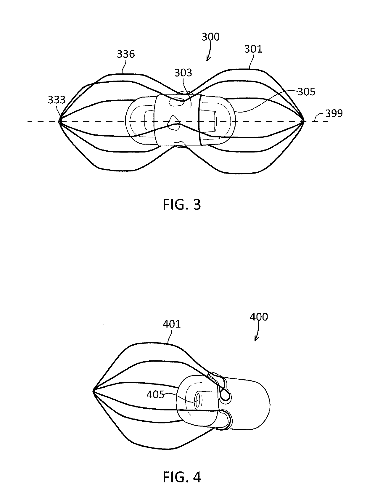
[0062]Described herein are devices for use with a capsule endoscope (CE) (a pill camera, pillcam, wireless capsule endoscope, or video capsule endoscope (VCE)) that significantly aid in more complete luminal visualization during capsule endoscopy. The devices create local distension of gastrointestinal luminal tissue away from the camera, improving diagnostic yield.

[0063]An exemplary luminal expansion device 300 is shown in FIG. 3. The device 300 includes a central attachment mechanism 303 configured to attach to the capsule endoscope 305. For example, the attachment mechanism 303 can be an annular ring. Further, the attachment mechanism 303 can be configured to attach by friction fit, adhesive, clamp, or other attachment mechanism to or around the capsule endoscope 305. The attachment mechanism 303 can be positioned, for example, around the central portion and / or end of the capsule endoscope 305 while still maintaining a clear lens.

[0064]A plurality of radiating struts 301 extend f...


PUM


Abstract

A device for distending a body lumen for enhanced visualization with a capsule endoscope that includes a camera lens includes an attachment element configured to attach to the capsule endoscope and a plurality of struts extending from the attachment element and meeting at an apex. The plurality of struts are configured to extend both axially and radially away from the camera lens so as to form a frame therearound.

Description

CROSS REFERENCE TO RELATED APPLICATIONS[0001]This application claims priority to U.S. Provisional Patent Application No. 62 / 376,816, filed Aug. 18, 2016 and titled “DEVICE AND METHOD FOR ENHANCED VISUALIZATION OF THE SMALL INTESTINE,” the entirety of which is incorporated by reference herein.INCORPORATION BY REFERENCE[0002]All publications and patent applications mentioned in this specification are herein incorporated by reference to the same extent as if each individual publication or patent application was specifically and individually indicated to be incorporated by reference.BACKGROUND[0003]Endoscopy, the study or examination of the inside of the body, is a common procedure performed by gastroenterologists inside of the gastrointestinal (GI) tract. It is typically accomplished with long, slender endoscopes, either rigid or flexible.[0004]For typical endoscopy, visualization of the GI tract is significantly enhanced by insufflation, i.e., the release of pressurized gas (air or CO...

Claims


Application Information

Patent Timeline
IPC IPC(8): A61B1/32A61B1/04A61B1/00
CPCA61B1/32A61B1/041A61B1/00101A61B1/00085A61B1/00148
Inventor TILSON, ALEXANDER Q.GOMES, GARRETT J.
Owner NEPTUNE MEDICAL INC
Learn More