In vitro method for identifying colorectal adenomas or colorectal cancer
a colorectal adenomas and in vitro technology, applied in the field of personalized medicine, can solve the problems of low compliance (less than 50%), the majority of said kind of patients can be wrongly classified as not having the disease, and the fit is not able to identify adenomas
- Summary
- Abstract
- Description
- Claims
- Application Information
AI Technical Summary
Benefits of technology
Problems solved by technology
Method used
Image
Examples
example 1
n of Study
[0049]A total of 300 subjects from eight Spanish hospitals (Hospital Clinic de Barcelona, Hospital de Burgos, Hospital de Vigo, Hospital de Tenerife, Hospital de Alicante, Hospital de Donosti, Hospital de Ourense and Hospital de Zaragoza) were prospectively included in this study: 193 patients newly diagnosed with sporadic colorectal neoplasia (92 with CRC and 101 with AA) and 100 healthy individuals without personal history of any cancer and with a recent colonoscopy confirming the lack of colorectal neoplastic lesions. Patients with AA were those with adenomas having a size of at least 10 mm or histologically having high grade dysplasia or >20% villous component. The characteristics of participants are shown in Table 1. Blood samples were collected prior to endoscopy or surgery in all individuals.
[0050]The study was approved by the Institutional Ethics Committee of Hospital Clinic of Barcelona (approval date: Mar. 11, 2014), and written informed consent was obtained from...
example 2
ction
[0051]Ten mL of whole blood from each participant were collected in EDTA K3 containing tubes. Blood samples were placed at 4° C. until plasma separation. Samples were centrifuged at 1,600×g for 10 min at 4° C. to spin down blood cells, and plasma was transferred into new tubes, followed by further centrifugation at 16,000×g for 10 minutes at 4° C. to completely remove cellular components. Plasma was then aliquoted and stored at −80° C. until use. miRNA isolation was performed using mirVana™ PARIS™ kit (Ambion by Life Technologies).
[0052]The miRNAs were extracted from all plasma samples, retro-transcribed, pre-amplified and analyzed by Real-Time quantitative PCR. For each RNA sample, 3 control miRNAs (cel-miR-39-3p as “spike-in”, and hsa-miR-1228-3p) and 5 candidate miRNAs (hsa-miR-15b-5p, hsa-miR-18a-5p, hsa-miR-29a-3p, hsa-miR-19a-3p and hsa-miR-19b-3p) were analyzed separately and in combination. Each miRNA in each sample was assessed in triplicate and mean Ct values were nor...
example 3
al Analysis
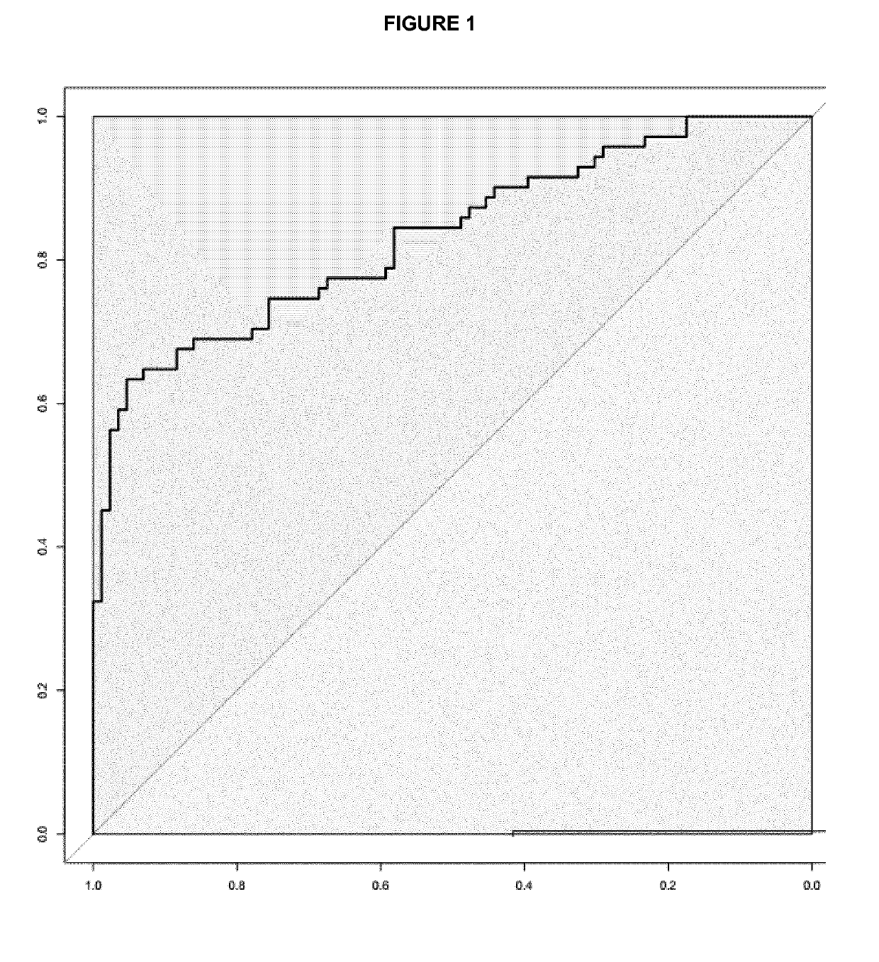
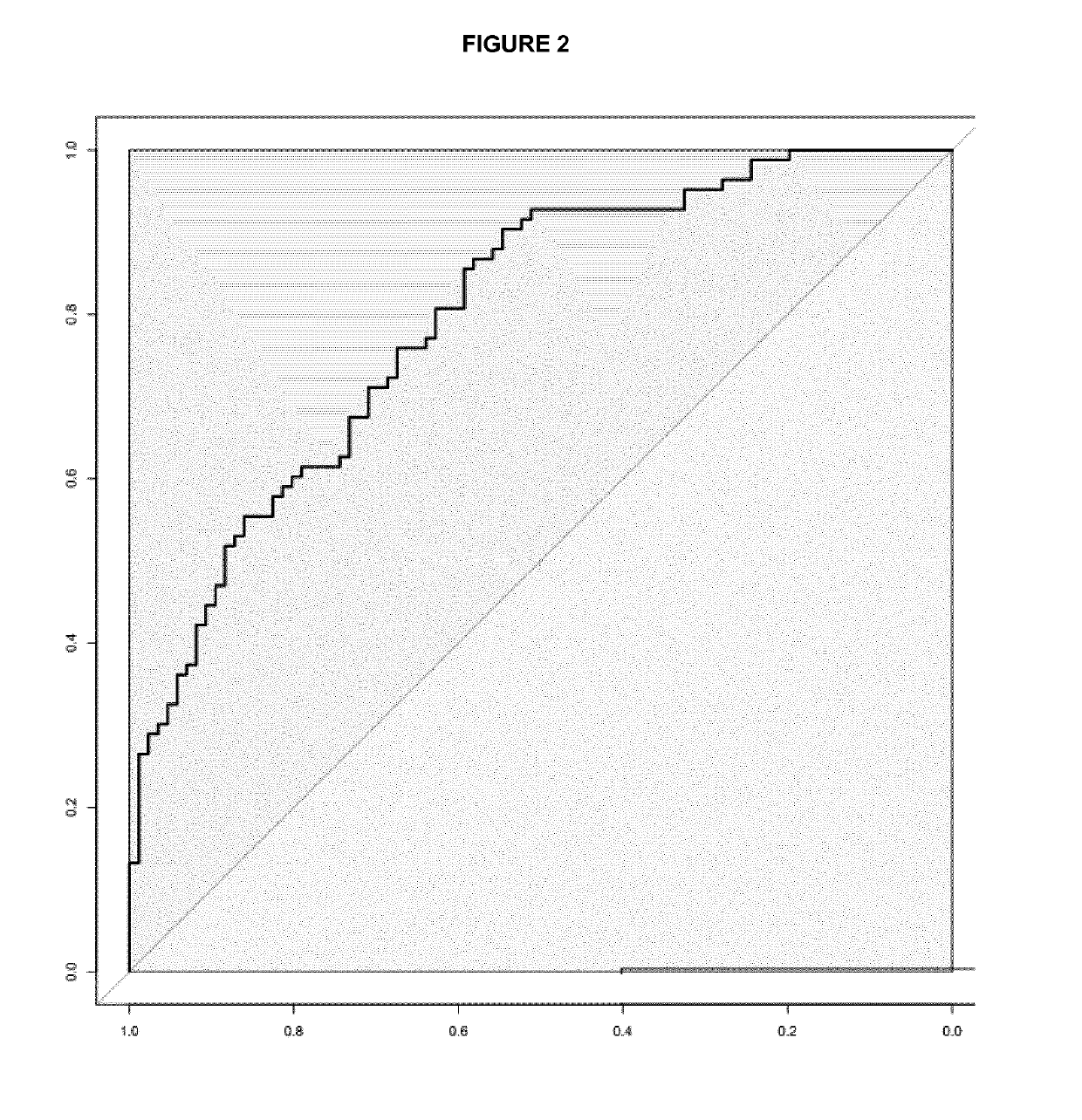
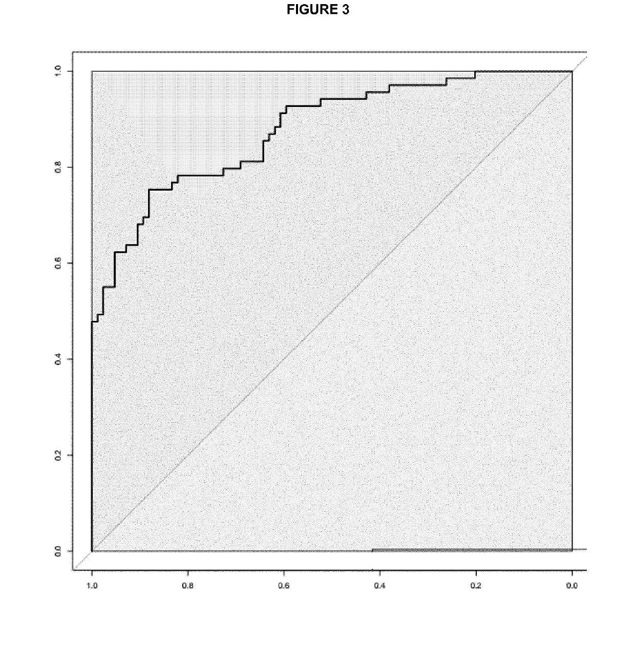
[0055]Differences in the miRNA expression levels (−DCt values) between different groups of patients were explored using multivariate logistic regression analysis adjusted by age, gender and site computing the odds ratio and intervals and their corresponding p values. ROC analysis plots and derived cut-points, as well as overall discriminative accuracy parameters, have been computed using DiagnosisMed R-package considering each miRNA expression as a continuous variable. Sensitivity (Sn), specificity (Sp), were calculated for several cut-points (the optimum cut-point associated with the minimum distance between the ROC curve and upper left corner, the cut-point associated with 80% Sn, with 85% Sn and with 90% Sn) (see Table 2 for colorectal cancer and Table 3 for advanced colorectal adenoma).
TABLE 2Colorectal CancerCriteraCut-offSnSpmiR-19a + miR-19b + miR-15bAUC = 0.845Min.ROC distance0.5307086AUC = 0.845Max.Youden Index0.6186494miR-19a + miR-29a + miR-15bAUC = 0.880Min.RO...
PUM
| Property | Measurement | Unit |
|---|---|---|
| Size | aaaaa | aaaaa |
| Fraction | aaaaa | aaaaa |
| Fraction | aaaaa | aaaaa |
Abstract
Description
Claims
Application Information
 - R&D
- Intellectual Property
- Life Sciences
- Materials
- Tech Scout
- Unparalleled Data Quality
- Higher Quality Content
- 60% Fewer Hallucinations
Browse by: Latest US Patents, China's latest patents, Technical Efficacy Thesaurus, Application Domain, Technology Topic, Popular Technical Reports.
© 2025 PatSnap. All rights reserved.Legal|Privacy policy|Modern Slavery Act Transparency Statement|Sitemap|About US| Contact US: help@patsnap.com


