
Ultrasound imaging system and method

Inactive Publication Date: 2019-10-31
GENERAL ELECTRIC CO
View PDF0 Cites 4 Cited by
  • Summary
  • Abstract
  • Description
  • Claims
  • Application Information

AI Technical Summary

Benefits of technology

This patent describes a method and system for acquiring and displaying ultrasound images of a person's body as a panoramic view. The system automatically divides the ultrasound image data into segments based on where it is acquired and can display video of the segments. This allows for a better understanding and analysis of the entire body of the person being examined.

Problems solved by technology

One issue with real-time imaging modalities is that operators may miss one or more items of interest in the image data during examination.
This can result in the operator missing one or more items of interest in the image data.
For example, imaging a relatively large organ, such as a lung, can be difficult in real-time imaging modalities such as ultrasound due to different parts of the organ being imaged and visible at different times. Additionally, the lung may be in near constant motion, with pathological items of interest (e.g., diseased, infected, or otherwise damaged areas) in different parts of the lung being visible at different times. As a result, the operator of the imaging system may not have the ability to see different moving parts of the lung at the same time, and can risk missing pathological items of interest.

Method used


Image

  • Ultrasound imaging system and method
  • Ultrasound imaging system and method
  • Ultrasound imaging system and method

Examples


Embodiment Construction

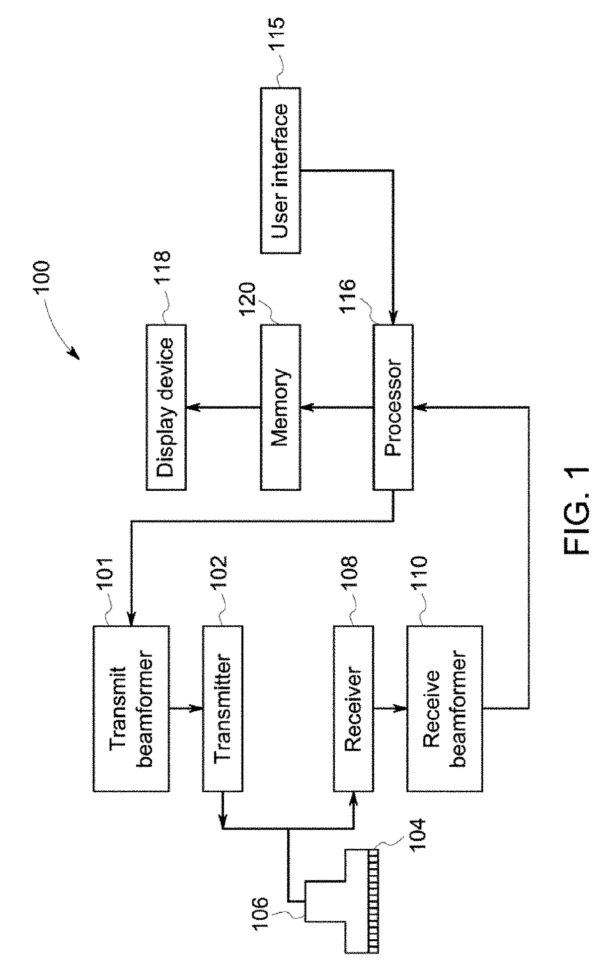
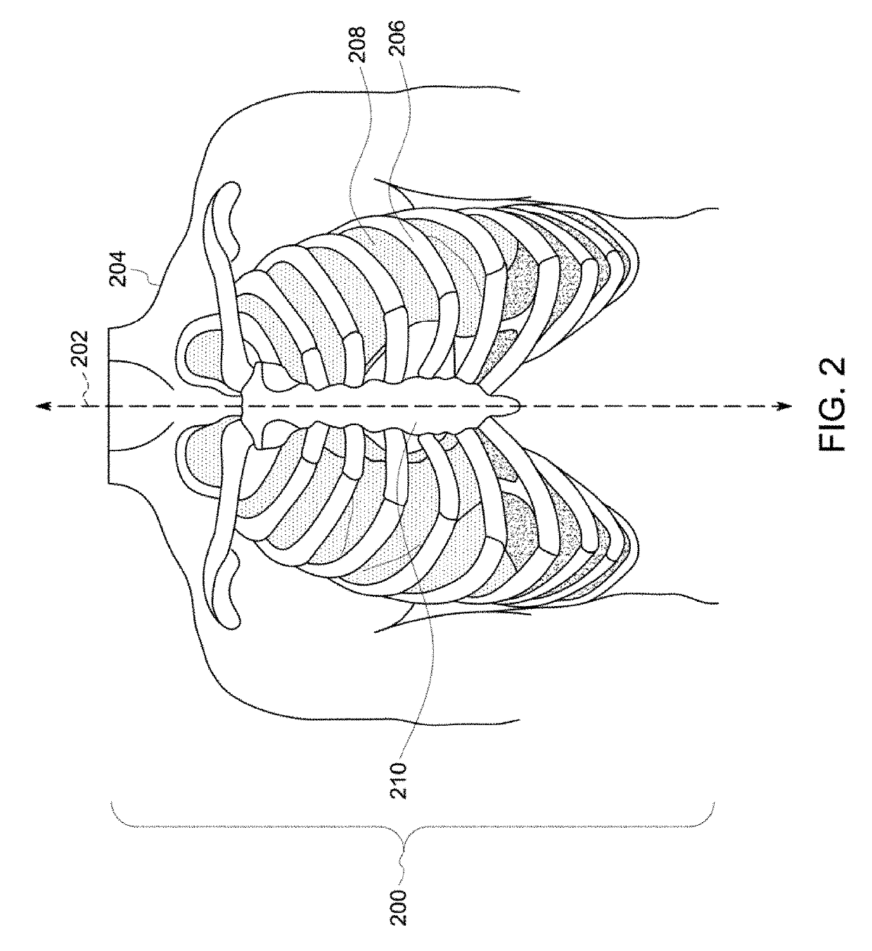
[0026]One or more embodiments of the inventive subject matter described herein provide imaging systems and methods that obtain real-time image data of a body and display a combined view of the image data representative of different portions of the body, with the combined view concurrently showing both dynamic and static image data. For example, the systems and methods can be used to image a body using ultrasound, and to present a panoramic view of the body with one or more portions of the body being shown with moving ultrasound image data (e.g., a video or cine) and one or more other portions of the same body being shown with static ultrasound image data (e.g., a still image). Alternatively, the combined view may show all dynamic image data. For example, the combined view may concurrently show dynamic image data of different intercostal areas of a person's lung. While the description herein focuses on the use of ultrasound image data and imaging lungs, not all embodiments are limite...


PUM


Abstract

An ultrasound imaging system and method acquires ultrasound image data from moving an ultrasound probe over a body of a person, automatically divides the ultrasound image data into segments of interest based on where the ultrasound image data was acquired, and displays a panoramic view of the ultrasound image data that includes two or more of the segments of interest with at least one of the segments of interest displayed as a video.

Description

FIELD[0001]The subject matter disclosed herein relates generally to ultrasound imaging systems.BACKGROUND[0002]Imaging systems generate image data representative of imaged bodies. Some imaging systems are not real-time diagnosis or examination modalities in that the image data from these types of systems is obtained, otherwise presented as images or videos at a later time (subsequent to acquisition of the image data), and then presented to an operator for examination.[0003]Other imaging systems are real-time diagnosis or examination modalities in that the image data from these types of systems is obtained and presented for diagnosis or examination by the operator in real-time. For example, the image data of a body can be visually presented to the operator for diagnosis or other examination while the imaging system continues obtaining additional image data of the same body.[0004]One issue with real-time imaging modalities is that operators may miss one or more items of interest in th...

Claims


Application Information

Patent Timeline
IPC IPC(8): A61B8/00G06T3/40A61B8/08
CPCA61B8/469G06T3/4038A61B8/463A61B8/523A61B8/5284G06T7/0012A61B8/4245A61B8/4444G06T2207/10132G06T2207/30061A61B8/085A61B8/4411A61B8/5223A61B8/481A61B8/08G06T2207/10016G06T2207/20021
Inventor HALMANN, MENACHEMOWEN, CYNTHIALYSYANSKY, PETERVARDI, MORSHIRAN, CARMIT
Owner GENERAL ELECTRIC CO
Learn More