

System for enhancing image quality of a subject using cbct (cone beam computed tomography)
- Summary
- Abstract
- Description
- Claims
- Application Information
AI Technical Summary
Benefits of technology
Problems solved by technology
Method used
Image

Examples
Embodiment Construction
Technical Field of the Invention
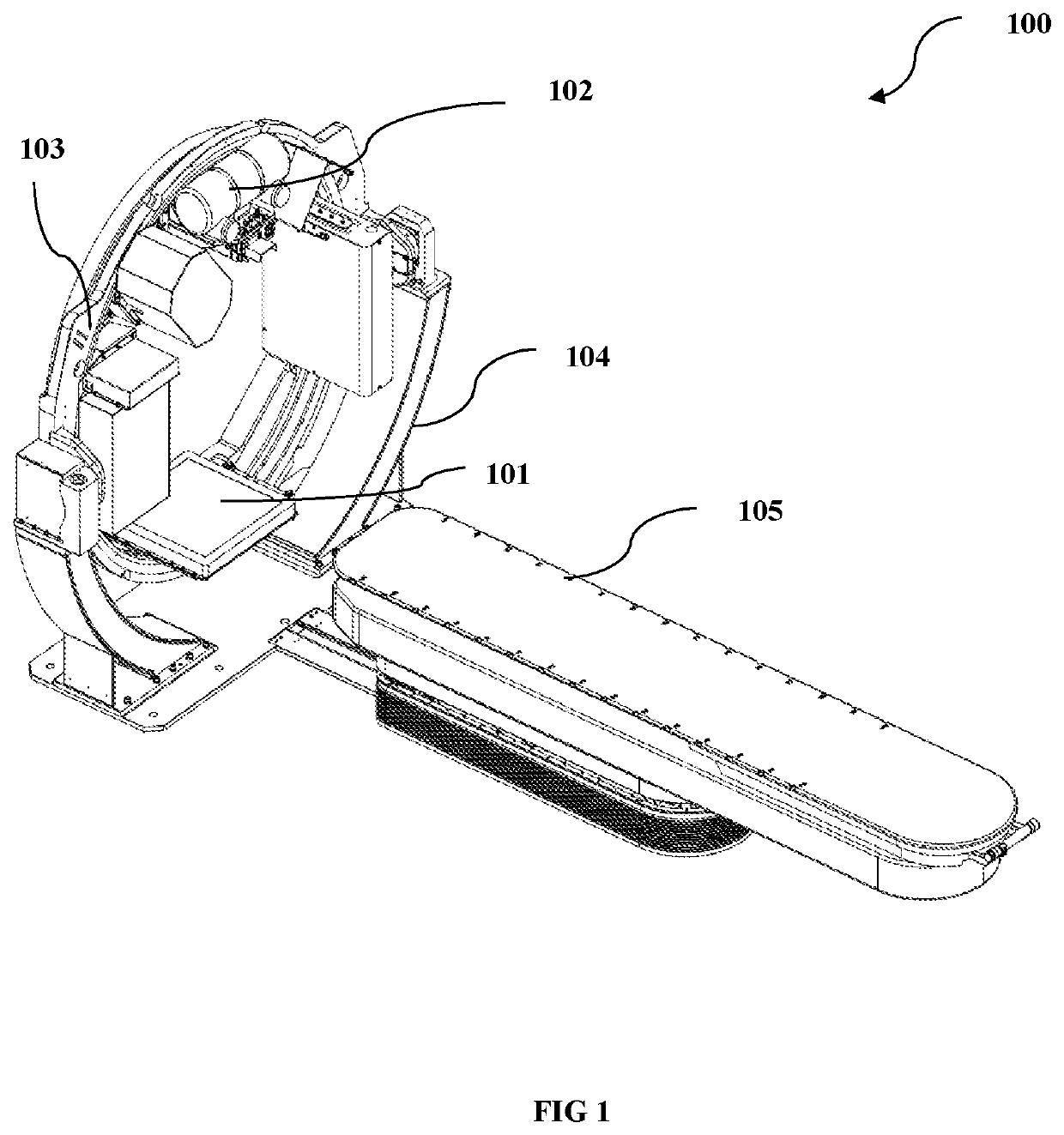
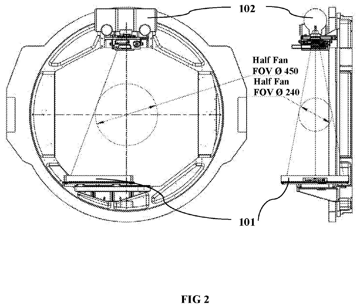
[0002]The present invention relates to the field of medical imaging techniques where x-ray source detector system is used to generate scan of entire region of interest using CBCT (Cone Beam Computed Tomography) technique.
Background of the Invention
[0003]Imaging systems are used in a wide variety of medical applications. Existing medical imaging systems rely on Conventional CT (Computed Tomography) devices or scanners in which multiple slices must be stacked to obtain a complete image. Conventional CT scanners employ the use of 1-D (one dimensional) detectors to generate an entire scan of the region of interest.
[0004]In another system, conventional fan beam spiral scan geometries are employed in x-ray use. Fan beam scans are obtained by transmission of the FOV (Field of View) by a narrow, fan-shaped beam of x-rays or multiple beams simultaneously. The x-ray beam generated by the tube is collimated to a fan shaped beam by rejecting the photons outside t...
PUM
 Abstract
Description
Claims
Application Information