A composite membrane comprising a decellularized amniotic membrane and a method for preparing the same

a technology of amniotic membrane and composite membrane, which is applied in the field of biomedical technology and composite membrane, can solve the problems of high cost associated with the preservation and delivery of fresh amniotic membrane, preventing its wide-spread use, and poor mechanical properties and fragile, and significant loss of some bioactive substances

Active Publication Date: 2020-06-11
WUHAN KANGCHUANG TECH +1
View PDF1 Cites 3 Cited by
  • Summary
  • Abstract
  • Description
  • Claims
  • Application Information

AI Technical Summary

Benefits of technology

[0091]The composite membrane of the invention has the advantages of both amniotic membrane and polymer fiber membrane, which retains the structure and biological function of an amniotic membrane, and also has the physical support function of the polymer fiber membrane. The composite membrane of the present application can be stored by freeze-drying to make ready-to-use products.
[0092]As compared with fresh or simply dehydrated amniotic membranes, the composite membrane of the present invention has good mechanical properties, is easy to operate, can be stored and transported at room temperature, reduces the cost of production, transportation and use, and therefore has a very good potential in applications. The composite membrane of the present invention can be used in a surgical operation, has good operability for a variety of operations including suturing, and will have a very wide range of applications in repair of a damaged soft tissue (for example, corneal injury, diabetic ulcers, arterial ulcers, lower extremity venous ulcers, or defects of tendons, muscles, or nerve tissue).
[0093]The method for preparing the composite membrane according to the present invention preserves the natural structure of stromal layer of amniotic membrane, and can make the decellularized amniotic membrane and the fibrous layer stably fit, so that the composite membrane has a stable integrated structure and excellent performance.

Problems solved by technology

The bioactivity and structure of amniotic membrane are best preserved in a freshly prepared amniotic membrane, but the high cost associated with the preservation and delivery of fresh amniotic membrane prevents its wide-spread use.
Lyophilized amniotic membranes have reduced the cost for preservation and transportation, but they are poor in mechanical properties and fragile.
However, these methods provide limited improvement, and may result in significant loss of some bioactive matters in amniotic membranes during the processing, and the introduced chemicals may potentially reduce the biocompatibility of amniotic membranes.
These approaches retain active substances of amniotic membranes to a certain extent, but completely destroy the natural structure of amniotic membrane, which has been confirmed to promote re-epithelialization.

Method used


Image

  • A composite membrane comprising a decellularized amniotic membrane and a method for preparing the same
  • A composite membrane comprising a decellularized amniotic membrane and a method for preparing the same
  • A composite membrane comprising a decellularized amniotic membrane and a method for preparing the same

Examples


example 1

Preparation and Treatment of PCL Nanofiber Membrane

[0120]1. Preparation of PCL nanofiber membrane: A solution of 12 wt % polycaprolactone (PCL, MW 70 kDa, Sigma-Aldrich) was prepared. A certain amount PCL particles were weighed and dissolved in a solution of dichloromethane: methanol=4:1 at a final concentration of 12%. The PCL solution was added to a syringe, and a 27 G syringe needle was mounted. The entire syringe was placed on a motor-driven syringe pump, and was propelled with a voltage of 12 kV at an injection speed of 2.5 mL / h. A grounded tin foil paper was used to receive the ejected PCL nanofibers to form a nanofiber membrane with a membrane thickness of 80 μm, and the average diameter of the PCL nanofibers was 517 nm (517±178 nm).

[0121]According to the above process, the PCL nanofiber membranes with thicknesses of 40 μm, 80 μm and 120 μm were prepared.

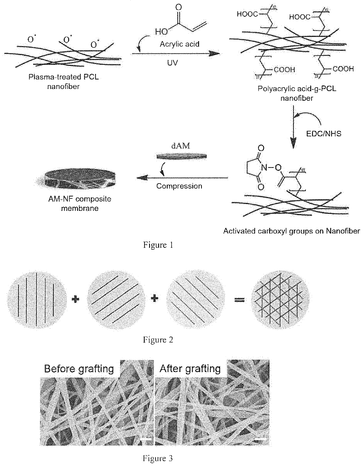
[0122]2. Surface treatment of PCL nanofiber membrane: the process was shown in FIG. 1.

[0123]1) The PCL nanofiber membrane w...

example 2

Decellularization Treatment of Amniotic Membrane

[0129]Fresh amniotic membrane was attached to a nitrocellulose membrane, with epithelial side up. A solution of 2.5% Dispase (Millipore) was prepared and dissolved in DMEM / F12 serum-free medium (Life Technology). The amniotic surface was covered with the enzyme solution, treated at 4° C. for 4 hours, and washed with PBS for 3 times. The amniotic membrane was placed in PBS solution and placed under a stereoscope. Epithelial cells were scraped off by using an Iris spatula from left to right, top to bottom. Under the stereoscope, it could be seen that white cell debris fell into PBS solution. The amniotic membrane was washed with PBS solution for 3 times.

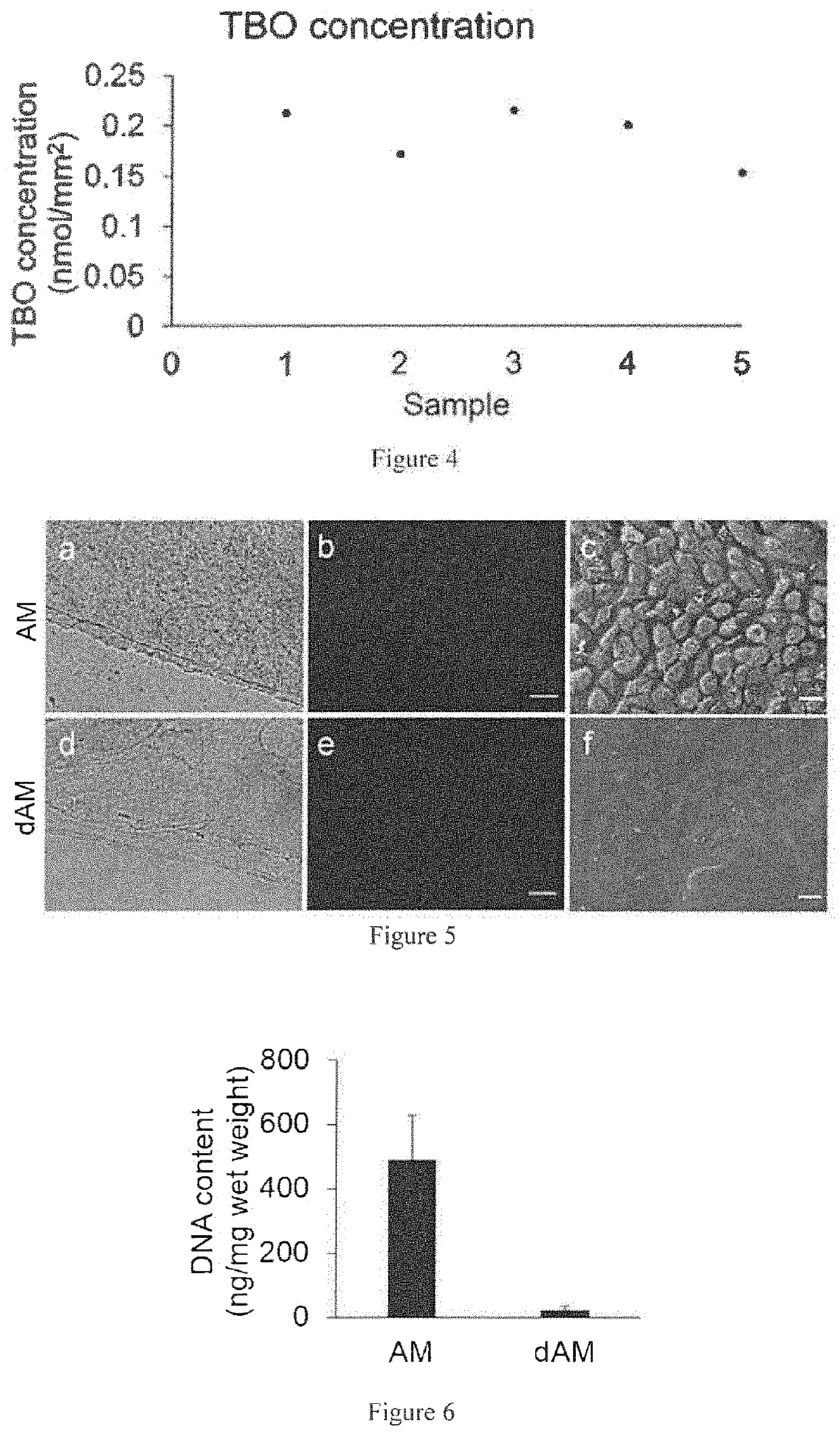
[0130]The thickness of the decellularized amniotic membrane was between 20 μm and 40 μm. The amniotic membrane was observed by optical microscope and scanning electron microscope before and after the decellularization, and the results were shown in FIG. 5 (scale bar: 20 μm). FIGS. 5a and ...

example 3

Preparation of Decellularized Amniotic Membrane-PCL Nanofiber Composite Membrane (“PCL-dAM Composite Membrane” For Abbreviation)

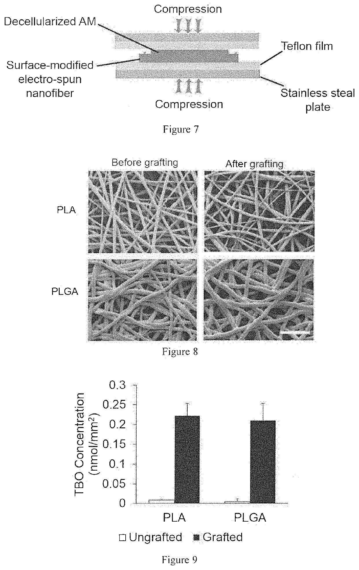
[0133]In an ultra-clean bench, the PCL nanofiber membrane was washed three times with PBS. The decellularized amniotic membrane with epithelial side down and stromal side up was spread out on the surface of Teflon (Polytetrafluoroethene, PTFE) film. The PCL nanofiber membrane was placed on the stromal side of the amniotic membrane. After that, another Teflon film was placed on the nanofiber membrane. The stacked decellularized amniotic membrane, the PCL nanofiber membrane and the Teflon films were placed between two stainless steel plates. The steel plates were placed in a vise, to which a compression of 5 to 7 MP was applied, and stayed at 4° C. for 12 hours. A condensation reaction was carried out between the carboxyl group on the surface of the PCL nanofiber membrane and the amino group on the surface of the decellularized amniotic membrane, and the reac...


PUM

PropertyMeasurementUnit
temperatureaaaaaaaaaa
diameteraaaaaaaaaa
diameteraaaaaaaaaa

Abstract

The present invention relates to the fled of biomedical technology, and relates to a composite membrane comprising a decellularized amniotic membrane, a use of the composite membrane, and a method for preparing the composite membrane.

Description

TECHNICAL FIELD[0001]The present invention relates to the field of biomedical technology, and relates to a composite membrane comprising a decellularized amniotic membrane, a use of the composite membrane and a method for preparing the composite membrane.BACKGROUND ART[0002]Amniotic membrane (AM) locates in the inner layer of placenta, is translucent, with soft texture, rich in a variety of growth factors and cytokines, and has been used in various clinical applications and animal studies for treatment and regeneration of soft tissues, such as in skin transplantation, ocular surface reconstruction, and treatment of diabetic ulcers, etc. The repair effects of amniotic membranes are mainly achieved via release of related factors, inhibition of inflammatory responses, promotion of re-epithelialization, inhibition of fibrosis and reduction of scar formation. Recent studies have shown that amniotic membranes can regulate inflammatory environment, promote transformation of M1 type macroph...

Claims


Application Information

Patent Timeline
Patent Type & Authority Applications(United States)
IPC IPC(8): A61L27/36A61L27/58A61L27/18C08L67/04A61L27/44A61L27/52
CPCA61L2430/40A61L27/52A61L27/3604A61L27/44A61L27/18C08L67/04A61L27/3691A61L2430/34A61L27/58A61L2400/12A61K35/50A61L27/50A61L27/56
Inventor LIU, HUANHUANWU, JUANBRAIN, GINNMAO, HAIQUAN
Owner WUHAN KANGCHUANG TECH
Why Eureka
Try Eureka