Methods, systems and devices for detecting inflammation
a technology of methods and systems, applied in the field of methods, systems and devices for the detection of inflammation, can solve the problems of poorly sensitive methods, and affecting the health system of developing countries, and limiting the application of methods
- Summary
- Abstract
- Description
- Claims
- Application Information
AI Technical Summary
Benefits of technology
Problems solved by technology
Method used
Image
Examples
example 1
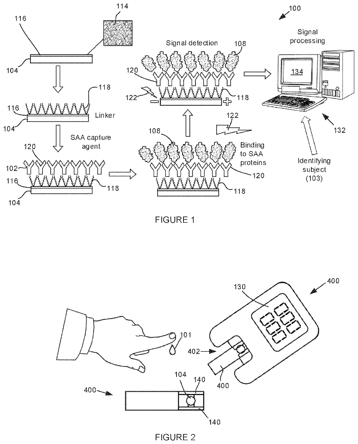
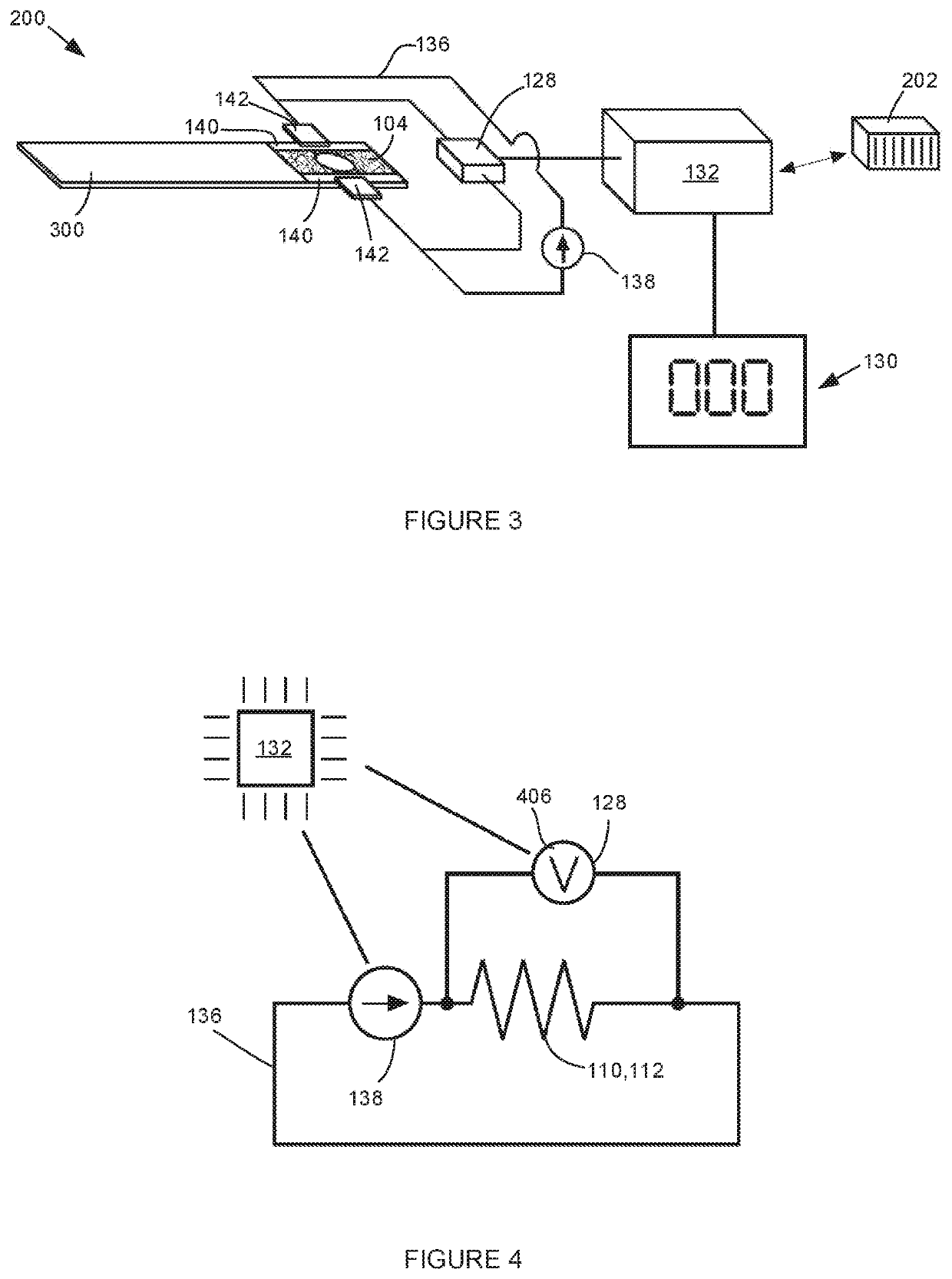
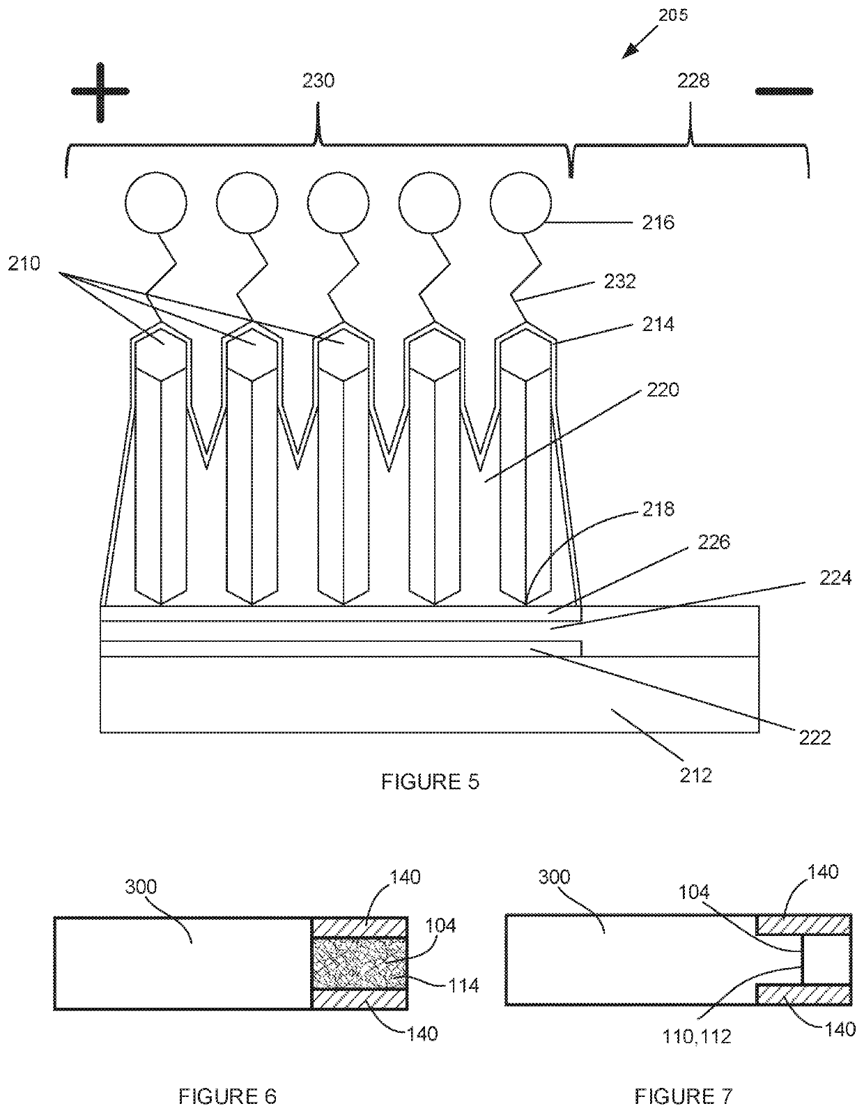
[0097]A system according to the invention includes an electrically conductive polymeric nanofibre for receiving a biological sample from a subject thereon. The nanofibre contains gold nanoparticles embedded therein and a linker, which may be a 3-mercaptopropanoic acid-containing SAM, securing SAA-binding antibodies or antibody fragments to the gold nanoparticles. In use, a constant current generator applies a constant current to the nanofibre and when SAA in the biological sample binds to the antibodies or antibody fragments resistance in the nanowire increases. The increase in resistance is proportional to the amount of SAA in the sample and can be detected by a detector, typically a volt meter or oscilloscope, as an impedance signal. The system further includes a processor in communication with the detector and configured to carry out the steps of: amplifying the resistance signal, converting the amplified signal to a digital signal, recording the digital signal, analysing the dig...
example 2
[0098]A test strip according to the invention includes an electrically conductive polymeric nanofibre for receiving a biological sample from a subject thereon. The nanofibre contains gold nanoparticles embedded therein and a linker, which may be a 3-mercaptopropanoic acid-containing SAM securing SAA-binding antibodies or antibody fragments to the gold nanoparticles. The test strip is configured to be positioned in a sample receiving zone of an inflammation measuring device in such a way that the nanofibre can be connected to a constant current generator. An increase in resistance in the nanowire resulting from binding of SAA to the antibody or antibody fragment is measurable by a resistance detector and a level of SAA in the sample determinable therefrom. A level of inflammation in the subject can then be assigned based on the level of SAA in the sample. The test strip is preferably manufactured to be a single-use, disposable test strip.
example 3
[0099]A device for use with the test strips of the second example is provided. The device includes a sample receiving zone for receiving the test strip and an electrical circuit to which the test strip is connectable when positioned in the sample receiving zone. The electrical circuit includes a constant current generator and a resistance detector, which is typically a volt meter or oscilloscope, for detecting resistance in the circuit. When the test strip is positioned in the sample receiving zone, a biological sample from a subject can be deposited on the test strip substrate and electrical resistance resulting from binding between SAA in the sample and the capture agent on the substrate detected. The device optionally includes a processor in communication with one or more of the constant current generator, resistance detector, or diode array detector for executing the steps of: amplifying the resistance signal, converting the amplified signal to a digital signal, recording the di...
PUM
 Abstract
Description
Claims
Application Information
 - R&D
- Intellectual Property
- Life Sciences
- Materials
- Tech Scout
- Unparalleled Data Quality
- Higher Quality Content
- 60% Fewer Hallucinations
Browse by: Latest US Patents, China's latest patents, Technical Efficacy Thesaurus, Application Domain, Technology Topic, Popular Technical Reports.
© 2025 PatSnap. All rights reserved.Legal|Privacy policy|Modern Slavery Act Transparency Statement|Sitemap|About US| Contact US: help@patsnap.com


