
A method to quantify the corneal parameters to improve biomechanical modeling

a biomechanical modeling and parameter technology, applied in medical simulation, medical informatics, medical automation diagnosis, etc., can solve the problems of corneal tomography change, lack of normal tensile strength of collagen fibrils, and poor organization of new collagen fibrils

Pending Publication Date: 2020-12-24
NARAYANA NETHRALAYA FOUND
View PDF1 Cites 2 Cited by
  • Summary
  • Abstract
  • Description
  • Claims
  • Application Information

AI Technical Summary

Benefits of technology

The patent text is about a method that helps predict how a cornea will behave after surgery. By analyzing the properties of the cornea before surgery, the method helps predict the long-term biomechanical response of the cornea after surgery. This can help improve the accuracy and effectiveness of corneal surgery.

Problems solved by technology

Further, the newly formed collagen fibrils are not well organized and lack the normal tensile strength of the collagen fibrils.
The surgery results in alteration of the biomechanical properties of the cornea resulting in change in tomography of the cornea as well.
However, the method is incomplete to predict the post-operative biomechanical changes in the corneal tissue.
However, the method fails to predict the post-operative biomechanical changes in the corneal tissue with patient variability.
However, the available methods disclose only the processes to analyse the quantitative changes.

Method used


Image

  • A method to quantify the corneal parameters to improve biomechanical modeling

Examples


Embodiment Construction

Technical Field of the Invention

[0001]The present invention relates to a method to quantify the human corneal tissue parameters to improve the biomechanical modelling of refractive and therapeutic procedures meant to reduce or eliminate refractive errors and unwanted wavefront aberrations.

BACKGROUND OF THE INVENTION

[0002]Cornea is the outermost layer of the eye, which is transparent, dome-shaped surface that covers the front part of the eye. Cornea receives nourishment from the tears and the aqueous humor that fills the chamber behind it due to absence of highly organized group of cells and blood vessels.

[0003]Cornea, the transparent anterior part of the eye covers the iris, pupil and an anterior chamber. Cornea together with the lens refracts light accounting for approximately two-thirds of the eye's total optical power.

[0004]Cornea has unmyelinated nerve endings, which are sensitive to stimuli, temperature and chemicals and causes an involuntary reflex to close the eyelid. The ref...


PUM


Abstract

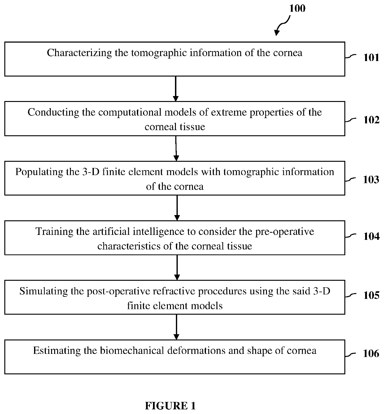
The invention relates to a method to quantify human corneal tissue parameters to improve biomechanical modelling of refractive and therapeutic procedures to reduce or eliminate refractive errors and unwanted wavefront aberrations. The analysis of the preoperative biomechanical properties of cornea assists in predicting the postoperative biomechanical response of cornea. The computational models of extreme properties of the corneal tissue are conducted and the 3-D finite element models are populated with tomographic information of the cornea and artificial intelligence is trained to consider the pre-operative characteristics of the corneal tissue. The post-operative refractive procedures are simulated using the said finite element models. Finally, biomechanical deformations are prospectively estimated and method builds a deep learning approach to link clinical features to changes in biochemical outcomes. The method predicts the post-operative biomechanics of the corneal tissue and derives long term postoperative biomechanical response of cornea.

Description

DESCRIPTION OF THE INVENTIONTechnical Field of the Invention[0001]The present invention relates to a method to quantify the human corneal tissue parameters to improve the biomechanical modelling of refractive and therapeutic procedures meant to reduce or eliminate refractive errors and unwanted wavefront aberrations.BACKGROUND OF THE INVENTION[0002]Cornea is the outermost layer of the eye, which is transparent, dome-shaped surface that covers the front part of the eye. Cornea receives nourishment from the tears and the aqueous humor that fills the chamber behind it due to absence of highly organized group of cells and blood vessels.[0003]Cornea, the transparent anterior part of the eye covers the iris, pupil and an anterior chamber. Cornea together with the lens refracts light accounting for approximately two-thirds of the eye's total optical power.[0004]Cornea has unmyelinated nerve endings, which are sensitive to stimuli, temperature and chemicals and causes an involuntary reflex ...

Claims


Application Information

Patent Timeline
Patent Type & Authority Applications(United States)
IPC IPC(8): A61B3/107A61B34/10G16H30/40G16H50/20G16H50/50
CPCG16H50/50G16H30/40A61B3/107A61B34/10A61B2034/105G16H50/20
Inventor ROY, ABHIJIT SINHASHETTY, ROHITFRANCIS, MATHEWCHANDAPURA, RACHANA
Owner NARAYANA NETHRALAYA FOUND
Learn More