Guided endotracheal intubation system

a technology of endotracheal intubation and guide tube, which is applied in the direction of respirator, medical science, surgery, etc., can solve the problems of death and disability, risk of accidental misplacement of endotracheal tube into esophagus, and serious brain damage or death, and achieves effective discrimination and control, and simple low-cost manufacturing

Inactive Publication Date: 2021-01-14
YISSUM RES DEV CO OF THE HEBREWUNIVERSITY OF JERUSALEM LTD +1
View PDF0 Cites 0 Cited by
  • Summary
  • Abstract
  • Description
  • Claims
  • Application Information

AI Technical Summary

Benefits of technology

[0007]The present disclosure describes new exemplary systems for the simple yet accurate viewing, guidance and execution of endotracheal intubation. The system utilizes a self-powered, stand-alone external illumination source, supplying a fixed output level of modulated illumination, applied externally to the region of the neck of the subject immediately external to the larynx region. The illumination source can be constructed in the form of a patch applied externally to the patient's throat region, and, because of its simple low-cost manufacture, could be a disposable component for single-event use with the instrument. This illumination is detected by an imaging system which receives its input from an endotracheal placement device, which can either have a detector array at its distal end, or can transmit the imaged view fiber-optically to a detector array disposed in the electronic sensing and control unit. The externally generated illumination is modulated, most conveniently by amplitude modulation, to enable the perceived or apparent imaged level to be adjusted by signal processing of the illumination detected internally within the subject's throat, and to enable effective discrimination and control of the external illumination penetrating into the trachea, as perceived by the sensing and control system, from the internal illumination provided by the endotracheal tube. The discrimination and level control can be achieved by using phase manipulation of the modulated illumination sensed, without the need for any inputs to the external illumination source at all, which itself provides a constant predetermined modulated output level of illumination.
[0008]This system enables the user to adjust the level of apparent tracheal illumination seen in the endoscopic images generated, the term apparent being used to emphasize that the actual tracheal illumination emitted from the trachea is constant (provided that the external illumination source is not moved, and that the patient does not move) and the different illumination level perceived is achieved by signal processing performed on the received image data. This system also enables the control system to use this perceived image data of the trachea to effectively perform automatic illumination level control, and automatic or semi-automatic steering of the endotracheal tube towards the trachea entrance, using images or image data having predetermined illumination and contrast characteristics.
[0009]The system enables the maintenance of the apparent imaged intensity of the light received from the external source at a level optimized for the detection of the trachea. If the level is too weak, the trachea may not be positively detectable, and if it is too strong, illumination may be collected from both the trachea and the esophagus, or light may be reflected or scattering from the surrounding tissues, thereby causing anatomical identification errors. Furthermore, as the location of the distal end of the endotracheal tube changes as it is advanced down the throat towards the vocal cords, or even into the trachea, the distance to the light source changes significantly, and the level of detected external illumination also changes.
[0011]The modulated illumination emitted by the external source should advantageously be at a wavelength which is readily transmitted through the tissues of the throat, such that it penetrates without undue attenuation, and also which has good detection sensitivity by commonly used photo-sensors, such as silicon-based CCD or CMOS arrays. Therefore, it is to be understood that use of the terms “light” or “illumination” in this disclosure is not intended to be limited to the visible region, but is understood to include any wavelength region which can thuswise be used by the system. Additionally, the modulation frequency should be commensurate with the frame frequency of readily available and standard video imaging devices, thereby keeping the system simple and of low cost.

Problems solved by technology

Failure to artificially ventilate an apneic patient rapidly could result in serious brain damage or death.
During patient intubation, there is a risk of accidental misplacement of the endotracheal tube into the esophagus.
This condition can in itself cause death and disability if not quickly detected.
However, when using internally illuminated endoscopic images, it is often difficult for the user to identify the exact trachea location in order to ensure performing the intubation properly, especially in trauma cases, where blood and secretions may be present, and where speed may be vital.
If the level is too weak, the trachea may not be positively detectable, and if it is too strong, illumination may be collected from both the trachea and the esophagus, or light may be reflected or scattering from the surrounding tissues, thereby causing anatomical identification errors.

Method used


Image

  • Guided endotracheal intubation system
  • Guided endotracheal intubation system
  • Guided endotracheal intubation system

Examples


Embodiment Construction

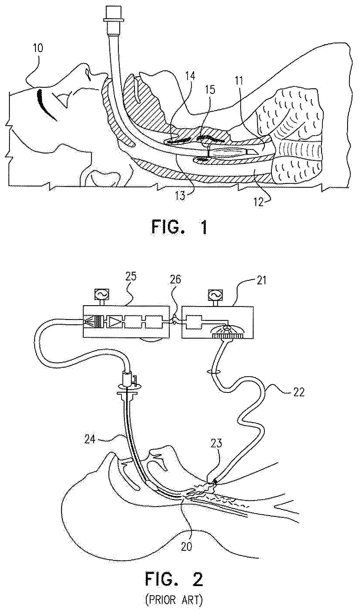
[0044]Reference is now made to FIG. 1, which illustrates schematically a conventional endotracheal procedure being performed on a patient 10. The trachea 11 is shown in its location in front of the esophagus 12, and an endotracheal intubation tube 13 has been successfully inserted past the epiglottis 14 and past the vocal chords 15 which are located at the junction of the trachea 11 and the esophagus 12, into the trachea. The problem of successfully negotiating the junction of the trachea and the esophagus is clear from FIG. 1. In commonly used procedures, the attending personnel manipulate the intubation tube into its correct position in the trachea by endoscopically viewing the progress of the distal tip of the intubation tube using illumination conveyed internally down the intubation tube assembly.

[0045]Reference is now made to FIG. 2, which is a schematic view of a prior art sensing system for tracheal intubation, as described in the above mentioned U.S. Pat. No. 5,560,351 to Gr...


PUM


Abstract

A guided tracheal intubation system using an autonomous modulated light source, outputting modulated illumination at a constant level, and externally applied to the subject's larynx region. An optical imaging system receives a video stream from within the subject's throat, including modulated illumination from the subject's trachea. A display control system performs signal processing on the modulated content of the images, and outputs frames of those images in which the intensity level of illumination from the trachea can be controlled without any need to change the illumination output from the modulated light source. The light source has no connection with the rest of the system, and need contain no more than a battery, a power supply circuit and a light source. It can therefore be of low cost and can be made disposable, such as in the form of an adhesive patch applied to the subject's neck.

Description

FIELD OF THE INVENTION[0001]The present invention relates to the field of endotracheal intubation, especially using external illumination to assist in the positioning of the endotracheal tube in the trachea of the subject.BACKGROUND OF THE INVENTION[0002]Endotracheal intubation is a well-known and widely used practice performed when normal ventilation of the patient's lungs may be impaired. Failure to artificially ventilate an apneic patient rapidly could result in serious brain damage or death. During patient intubation, a flexible tube, also known as an endotracheal tube, is used. A distal end of the tube is placed within the patient's trachea. The proximal end of the tube can be attached to a resuscitator bag or any other device, supporting the respiratory process. During patient intubation, there is a risk of accidental misplacement of the endotracheal tube into the esophagus. This condition can in itself cause death and disability if not quickly detected. When performing trache...

Claims


Application Information

Patent Timeline
Patent Type & Authority Applications(United States)
IPC IPC(8): A61B1/06A61B1/00A61B1/267A61M16/04A61B1/04
CPCA61B1/06A61B1/00009A61B1/04A61M16/0488A61B1/0661A61B1/267A61B1/00165A61B1/2676A61B2560/06A61B2576/02A61M2210/1032A61B1/00097A61B1/0655
Inventor HAYUT, ITAIFRIED, ELCHANANNAHMIAS, YAAKOVWEISS-SADAN, TOMMYSHREM, ARIEL
Owner YISSUM RES DEV CO OF THE HEBREWUNIVERSITY OF JERUSALEM LTD
Why Eureka
Try Eureka