
Eye surgery surgical system and computer implemented method for providing the position of at least one trocar point

a surgical system and computer-implemented technology, applied in the field of eye surgery surgical system and computer-implemented, can solve the problems of hollow needle not reaching the lumen of the vessel, impairing the surgeon's view of the fundus during an eye operation, hollow needle slipping out of the vessel, etc., and achieve the effect of optimizing workflows

Pending Publication Date: 2021-03-04
CARL ZEISS MEDITEC AG
View PDF0 Cites 3 Cited by
  • Summary
  • Abstract
  • Description
  • Claims
  • Application Information

AI Technical Summary

Benefits of technology

The invention is a system and method for eye surgery that helps to accurately position a tool (trocar) on the eye's sclera for a surgical intervention. The system includes a device that references the patient's eye model to the actual eye, making it easier to visualize the correct position of the trocar on the eye. The trocar point calculation routine takes into account the surgeon's posture and maximizes the distance of the instruments from critical tissue in the eye model to reduce the risk of complications. This results in an optimized workflow for eye surgery.

Problems solved by technology

If the angle of attack is too flat, the hollow needle cannot reach the lumen of the vessel.
Then, even very small relative movements between hollow needle and fundus can lead to the hollow needle piercing through the vessel or slipping out of the vessel again.
Blood that has entered the vitreous humour can impair the surgeon's view of the fundus during an eye operation.
A possible consequence is that the retinal vessel can no longer be treated correctly.
This is because the angle of attack for a surgical tool of the surgical instrument with respect to a structure in the interior of the patient's eye, for example, a pointed cannula, a blade or a pair of scissors, can subsequently no longer be varied as desired.
In the case of surgical operations in the interior of a patient's eye, a surgeon often handles surgical instruments with a surgical tool at angles of attack with respect to structures in the interior of the patient's eye which are very inexpedient and therefore could cause complications.
Often, it only becomes clear after placing the trocar that a planned intervention on the retina cannot be carried out because the surgical instrument introduced into the interior of the patient's eye through the trocar does not allow the angles of attack of the surgical tool of the surgical instrument required for the intervention.
However, inserting and removing different surgical instruments through a placed trocar to find out which surgical instrument allows the surgical intervention to be carried out best lengthens the time required for the surgical intervention to be carried out.
Moreover, keeping available and using different surgical instruments causes significant costs during procurement and operation.
However, such surgical instruments are expensive to produce and can only be sterilized with great outlay, if at all.
If it turns out that it is not possible to carry out the planned surgical intervention in the interior of the patient's eye using the placed trocar, it is necessary to place the trocar anew.
Particularly if use is made of further surgical instruments and auxiliary devices in the form of stands or robots, this requires a lot of time since the further surgical instruments may also have to be repositioned and, for instance, robots need to be realigned and programmed for the new so-called remote center of motion (RCM).
Then, a patient inevitably experiences additional injury during the operation and the costs for carrying out the surgical intervention increase on account of the need for additional consumables, required in that state, and the lengthening of the duration of the operation.

Method used


Image

  • Eye surgery surgical system and computer implemented method for providing the position of at least one trocar point
  • Eye surgery surgical system and computer implemented method for providing the position of at least one trocar point
  • Eye surgery surgical system and computer implemented method for providing the position of at least one trocar point

Examples


Embodiment Construction

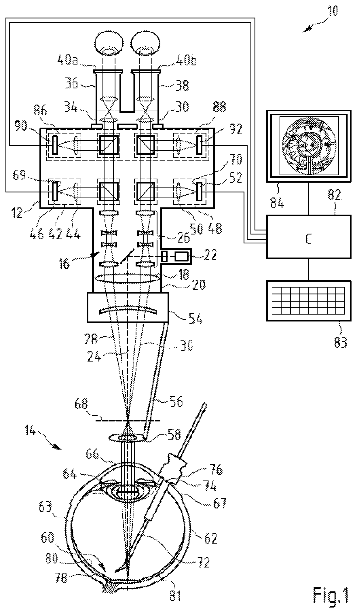
[0038]The eye surgery surgical system 10 shown in FIG. 1 contains a surgical microscope 12, which serves for the stereoscopic observation of an object region 14. The surgical microscope 12 comprises an imaging optical unit 16 with a microscope main objective system 18, the imaging optical unit being received in a main body 20. In the surgical microscope 12, there is an illumination device 22, which facilitates the illumination of the object region 14 with an illumination beam path 24, which passes through the microscope main objective system 18. The surgical microscope 12 has an afocal magnification system 26, through which a first stereoscopic partial observation beam path 28 and a second stereoscopic partial observation beam path 30 are guided. The surgical microscope 12 has a binocular tube 34 connected to an interface 32 of the main body 20, the binocular tube having a first eyepiece 36 and a second eyepiece 38 for a left and a right eye 40a, 40b of a surgeon. The microscope mai...


PUM


Abstract

An eye surgery surgical system includes a visualization device for visualizing the position of at least one trocar point for a trocar on the sclera of a patient's eye. The trocar serves to introduce a surgical instrument configured for surgical interventions on the fundus of the patient's eye into the patient's eye. The eye surgery surgical system also includes a computer unit which is configured to provide the position of the at least one trocar point to the visualization device. Here, the computer unit contains a trocar point computation routine configured to calculate the position of the at least one trocar point from the location of at least one surgical site in a model of the patient's eye and from geometric data relating to at least one surgical instrument introducible into the patient's eye through the trocar.

Description

CROSS REFERENCE TO RELATED APPLICATION[0001]This application claims priority of German patent application no. 10 2019 123 742.4, filed Sep. 4, 2019, the entire content of which is incorporated herein by reference.FIELD OF THE INVENTION[0002]The invention relates to an eye surgery surgical system and a computer-implemented method for providing the position of at least one trocar point for a trocar on the sclera of a patient's eye, which serves to introduce at least one surgical instrument configured for surgical interventions on the fundus of the patient's eye into the patient's eye. Moreover, the invention relates to a computer program.BACKGROUND OF THE INVENTION[0003]Vitreoretinal surgery uses surgical instruments that are guided into the interior of a patient's eye through a trocar. By way of example, to carry out so-called retinal vein cannulation (RVC), cannulas with a shaft body are used, at the end of which a surgical tool in the form of a fine hollow needle, as a rule angled ...

Claims


Application Information

Patent Timeline
IPC IPC(8): A61F9/007A61B3/00A61B3/13A61B3/12
CPCA61F9/007A61B3/0008A61B3/12A61B3/0025A61B3/13A61B3/0041A61F9/00736A61B17/3421A61B34/10
Inventor VOIGT, CHRISTIANANDREWS, DELBERT PETERHECKER-DENSCHLAG, NANCYMATZ, HOLGERHAUGER, CHRISTOPH
Owner CARL ZEISS MEDITEC AG
Learn More