

Image-based device tracking
a technology of image-based devices and tracking devices, which is applied in the direction of radiation diagnostic clinical applications, catheters, applications, etc., can solve the problems of difficult to recognize the device in an ultrasound image, difficult to properly interpret the image, and difficult to get an optimal view for guidance and treatmen
- Summary
- Abstract
- Description
- Claims
- Application Information
AI Technical Summary
Benefits of technology
Problems solved by technology
Method used
Image

Examples
Embodiment Construction
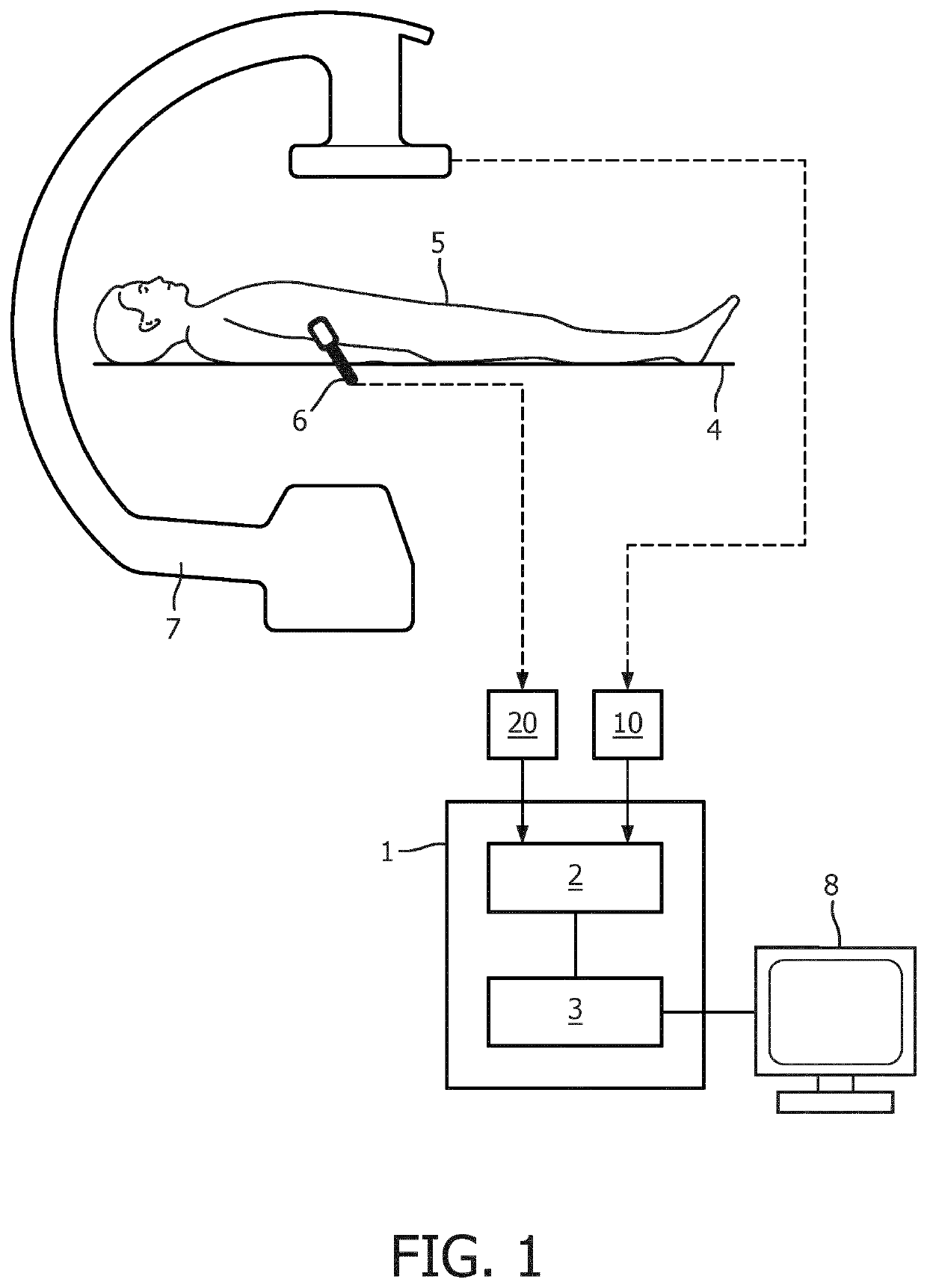
[0054]FIG. 1 shows an exemplary apparatus 1 for supporting a medical procedure as described herein. The apparatus comprises an input unit 2 for receiving first and second image data, for example X-ray medical image data from a C-arm imaging device 7 and ultrasound medical image data from an ultrasound probe 6. The image data are being acquired during a procedure being carried out on patient 5 lying on support 4. A processing unit 3 is provided for processing the image data and identifying a device of interest, in accordance with the examples further described herein.
[0055]Optionally, the apparatus is part of an imaging system, for example an ultrasound imaging system further comprising an ultrasound probe 6. In this case, for example, the apparatus may be configured as, or form part of, an ultrasound console. Alternatively, a computer program product may be provided to be executed by a processing unit of an ultrasound console.
[0056]Alternatively, the apparatus may be part of a C-arm...
PUM
 Abstract
Description
Claims
Application Information