

Ultrasound visual protocols
a visual protocol and ultrasound technology, applied in the field of ultrasound visual protocols, can solve the problems of inefficiency of any particular user's use of ultrasound protocol information, time-consuming, and complex review of completed ultrasound protocols
- Summary
- Abstract
- Description
- Claims
- Application Information
AI Technical Summary
Benefits of technology
Problems solved by technology
Method used
Image

Examples
Embodiment Construction
Overview
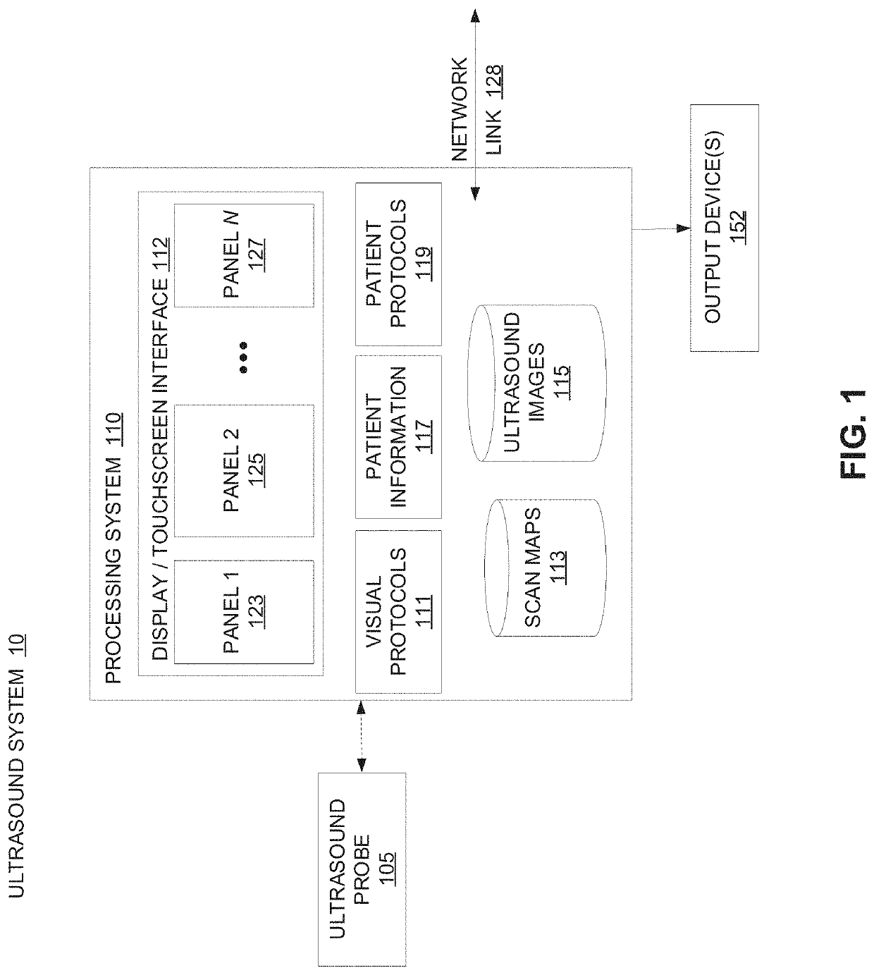
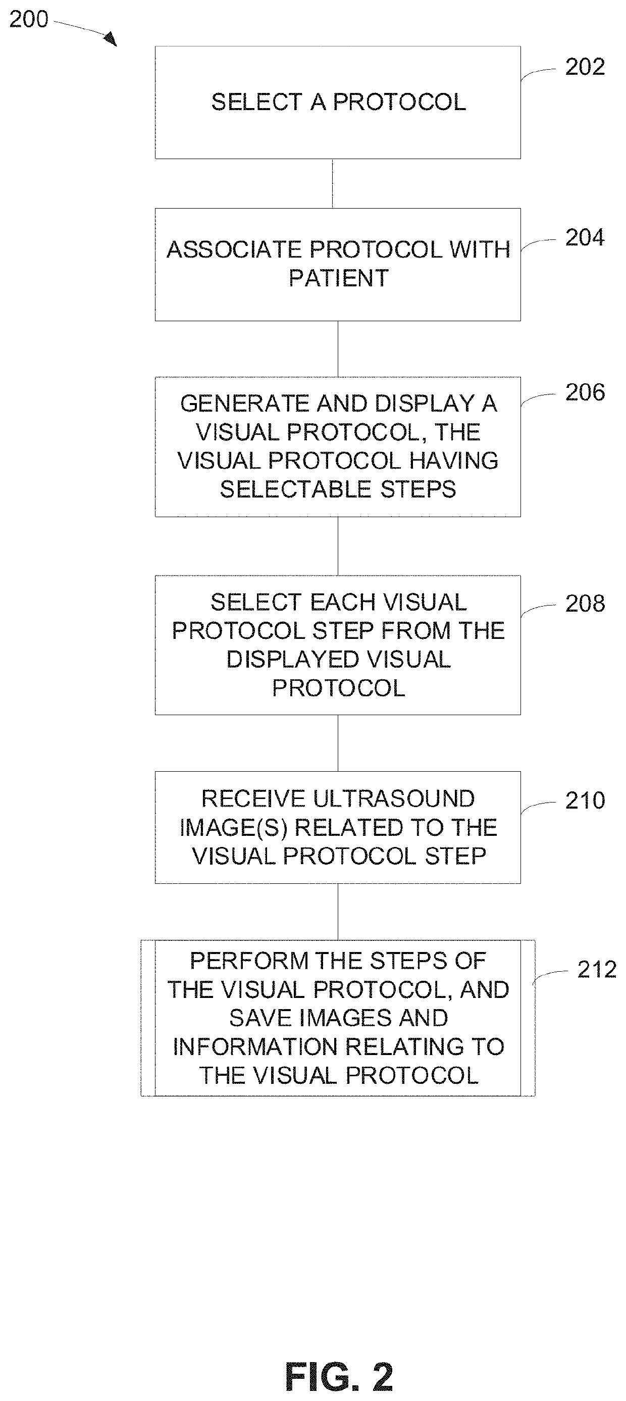
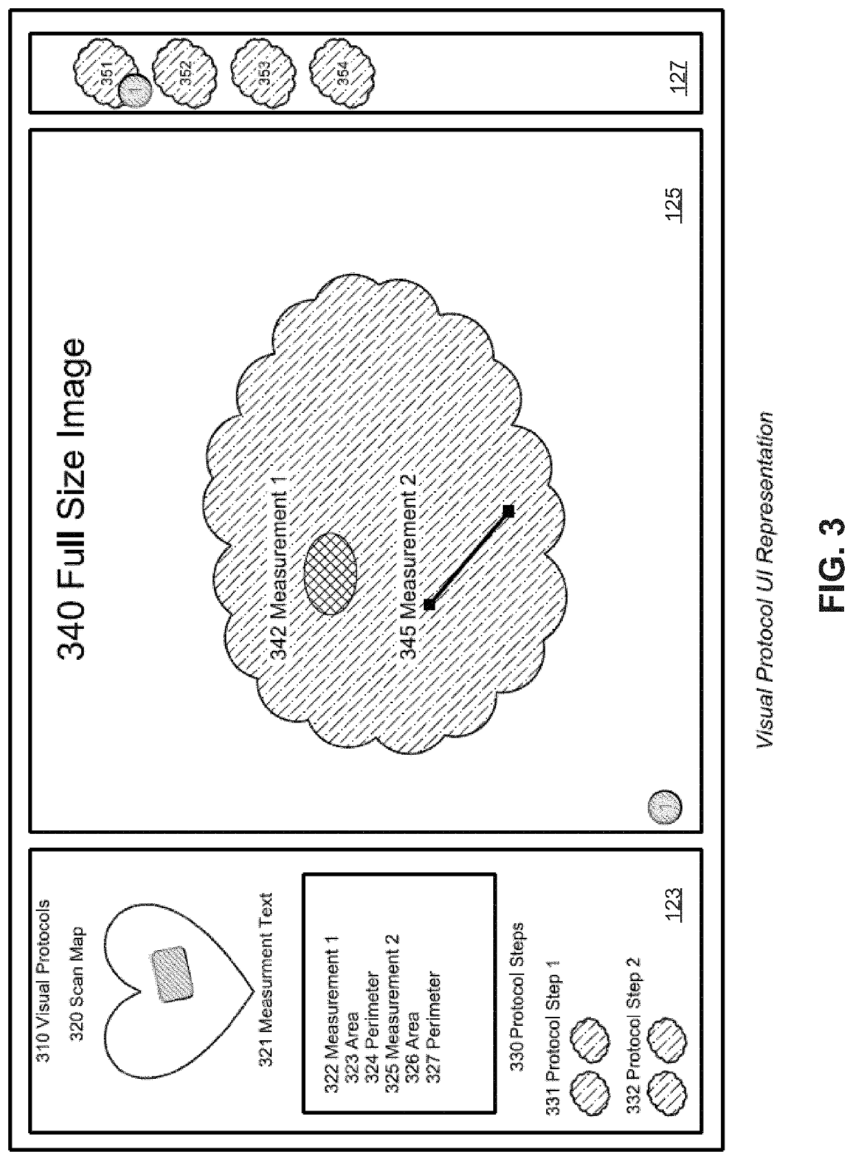
[0016]Embodiments of systems and methods for performing visual ultrasound protocols are disclosed herein. A software application provides a user interface for selecting, visualizing, and interacting with visual ultrasound protocols, including ultrasound image association, annotation and measurement of objects in an ultrasound image, including suspect objects and a patient's anatomy in an image. For example, a method may include selecting a protocol, from a list display protocols, to perform for patient. The method may also include selecting a protocol step, saving captured ultrasound images of interest in associating them with a protocol step, adding information (e.g., a scan position) to a scan map that depicts a graphical representation of the scanned anatomy, and adding annotations and / or measurements to the protocol.
[0017]Each protocol step may be linked to ultrasound images and other information relating to the protocol step, such that when the protocol step later is se...
PUM
 Abstract
Description
Claims
Application Information