Fundus photographic technique to determine eye refraction for optic disc size calculations

a technology of optic disc and calculation method, which is applied in the field ofophthamological retinal imaging, can solve the problems of inability to easily put into everyday practice, cumbersome measurement methods, and differences in image magnification of fundus photo, and achieves rapid glaucoma screening, important socioeconomic benefits, and reduce patient disability

Inactive Publication Date: 2005-02-15
QUIGLEY MICHAEL
View PDF9 Cites 5 Cited by
  • Summary
  • Abstract
  • Description
  • Claims
  • Application Information

AI Technical Summary

Benefits of technology

It is a goal of the present invention to provide a simple means to perform the mentioned magnification calculation to permit the determination of true sizes of optic nerve and optic nerve structures in order to allow for rapid screening for glaucoma from a simple fundus photograph. Early diagnosis of glaucoma would decrease patient disability and hence have important socioeconomic benefits.

Problems solved by technology

In ophthalmic imaging, the determination of scale is a challenge for the ophthalmologist using a fundus camera.
These techniques are tedious as they require photograph and supplemental testing either to determine the axial length of the eye or its optical error (glass refraction) or its optical error and the curvature of the cornea.
However, as alluded to above, not all eyes are the same in terms of their optical errors and sizes which results in differences in image magnification on the fundus photo.
This can be impractical.
While feasible, such measurement methods are cumbersome and not easily put into everyday practice.

Method used


Image

  • Fundus photographic technique to determine eye refraction for optic disc size calculations
  • Fundus photographic technique to determine eye refraction for optic disc size calculations

Examples


Embodiment Construction

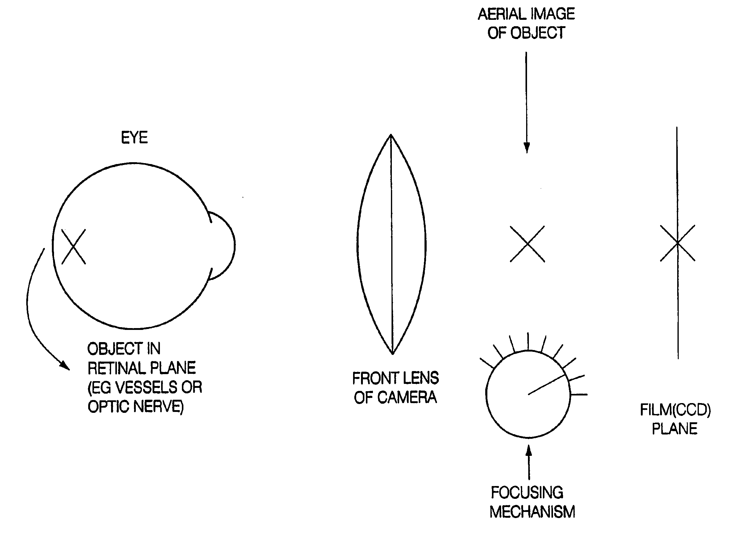
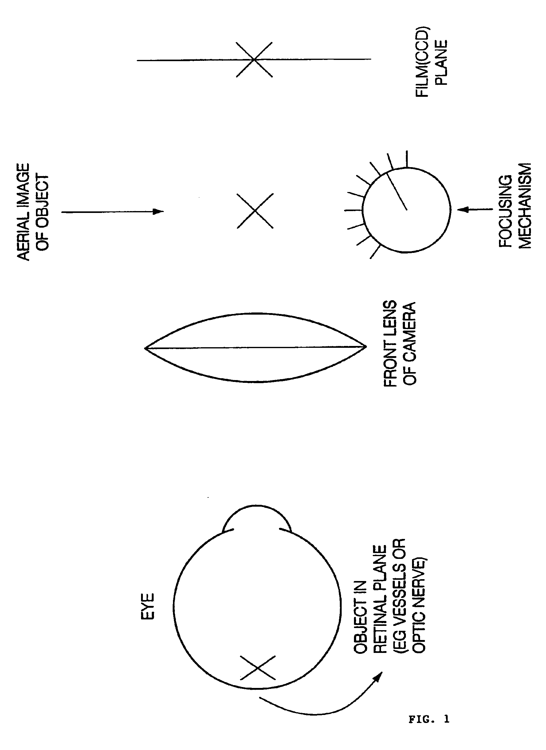
According to the invention, the position of the focusing knob or mechanism on the fundus camera is recorded, which position reflects the optical error or glass refraction of the eye. The glass refraction of the eye is used to calculate the object to image size ratio or magnification. This focus mechanism position is preferably automatically incorporated into a means to calculate the eye-camera magnification to arrive at a good estimate of the true or absolute measurements of retinal structures on the fundus photograph, such as the optic nerve, its components including the neuroretinal rim area and the cup. All of these measurements are important in the diagnosis of glaucoma.

The Applicant of the present application has noted that the observation and photography systems of the fundus camera rely on the principle of indirect ophthalmoscopy (“Some Essential Optical features of the Zeiss Fundus Camera”, Bengtsson B and Krakau C. E. T. Acta Ophthalmologica Vol 55, 1977:123-131). The locat...


PUM


Abstract

The determination of magnification in an ophthalmic fundus camera image is achieved by detecting the focusing mechanism position, and using a calibration function to calculate the magnification from the focus position.

Description

FIELD OF THE INVENTIONThe present invention relates generally to ophthamological retinal imaging, and in particular to a method and apparatus for determining an object size, such as the optic disc or other retinal object, in an image from a fundus camera.BACKGROUND OF THE INVENTIONIn ophthalmic imaging, the determination of scale is a challenge for the ophthalmologist using a fundus camera.For example, the determination of the true size of the optic nerve head from its image on a fundus photograph is of great clinical importance in the diagnosis of glaucoma. As is well recognized in the art, glaucoma is a visually debilitating disease process resulting in damage to the nerve tissue of the optic nerve and having prevalence in the over 40 age group of about 1% (see Duane's Clinical Ophthalmology, Tasman W and Jaeger E editors, Vol. 3, Chapter 42, Glaucoma: general concepts).The current methods for determining optic disc size include performing an ultrasound of the eye to determine the...

Claims


Application Information

Patent Timeline
Patent Type & Authority Patents(United States)
IPC IPC(8): A61B3/12
CPCA61B3/12
Inventor QUIGLEY, MICHAEL
Owner QUIGLEY MICHAEL
Why Eureka
Try Eureka