Arthroscopic impedance probe to detect cartilage degeneration

a technology of impedance probe and arthroscopic probe, which is applied in the field of non-destructive arthroscopic diagnostic probes, can solve problems such as voltage difference between electrodes, and achieve the effects of resisting tension and shear loading, resisting compressive loading, and high swelling pressur

Inactive Publication Date: 2005-04-19
MASSACHUSETTS INST OF TECH
View PDF6 Cites 6 Cited by
  • Summary
  • Abstract
  • Description
  • Claims
  • Application Information

AI Technical Summary

Problems solved by technology

When current is applied to the tissue there is a distribution of current density and an electric field within the tissue, and a resulting voltage difference across the electrodes applying the current.

Method used


Image

  • Arthroscopic impedance probe to detect cartilage degeneration
  • Arthroscopic impedance probe to detect cartilage degeneration
  • Arthroscopic impedance probe to detect cartilage degeneration

Examples


Embodiment Construction

Operation of the Invention

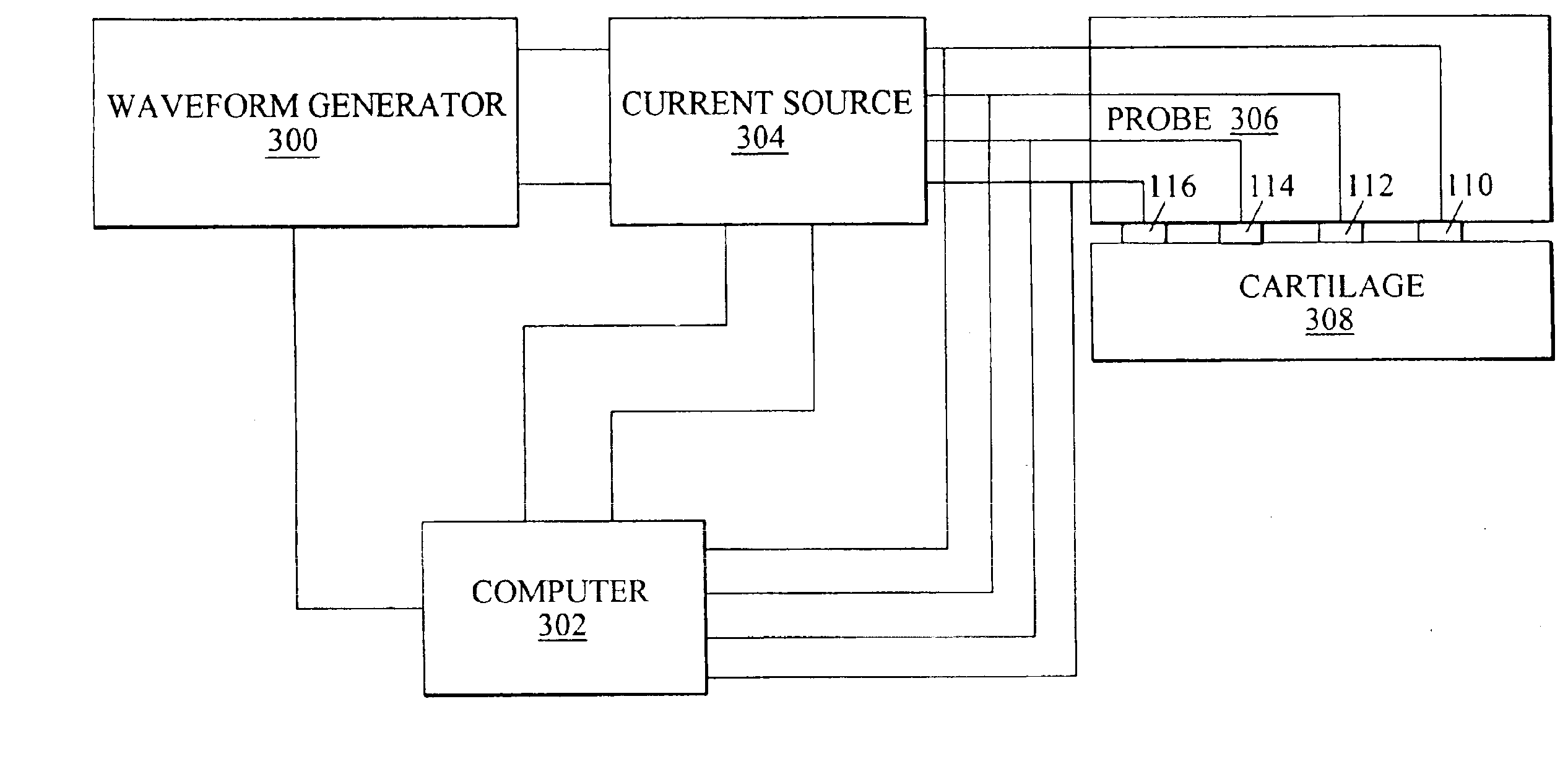
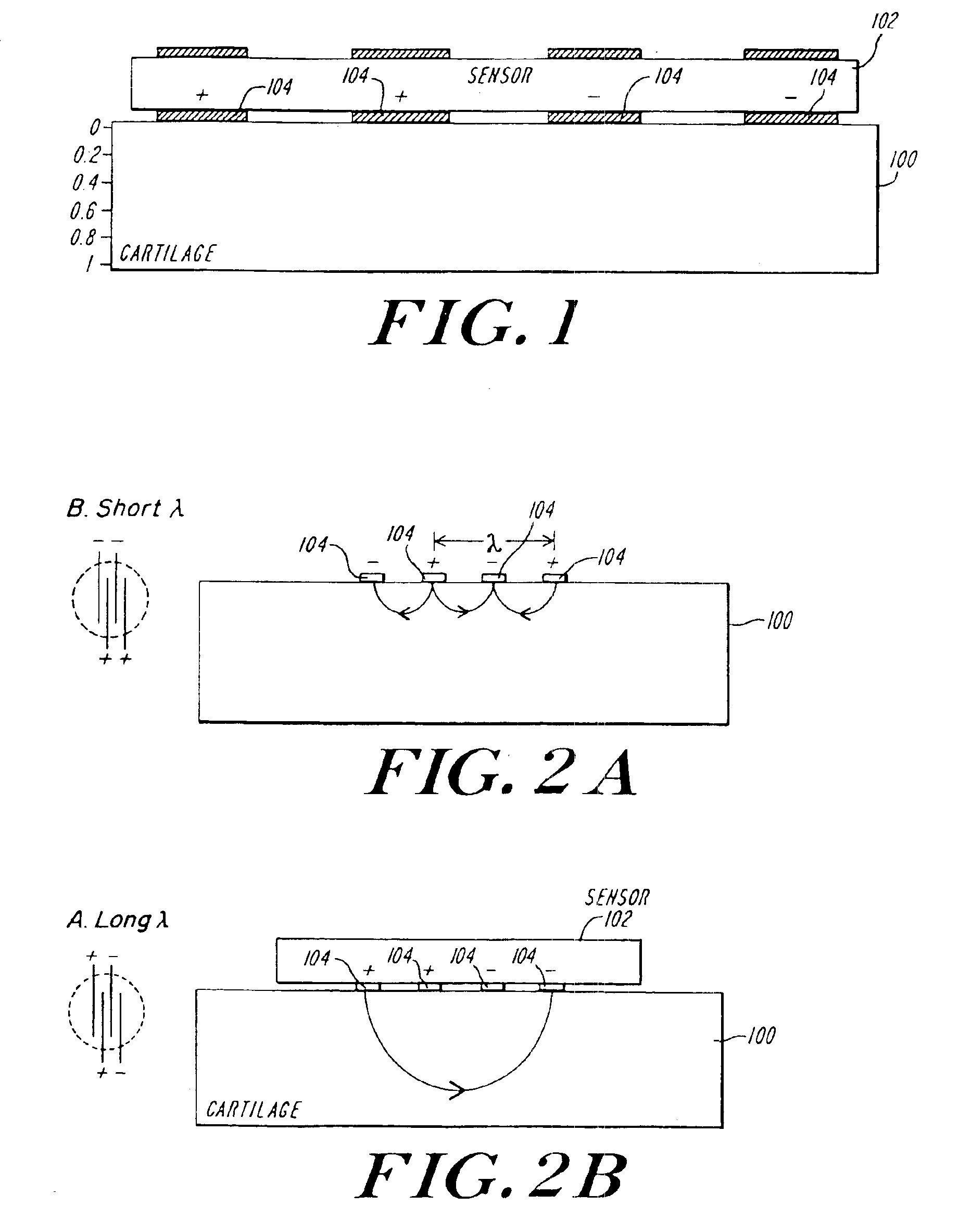
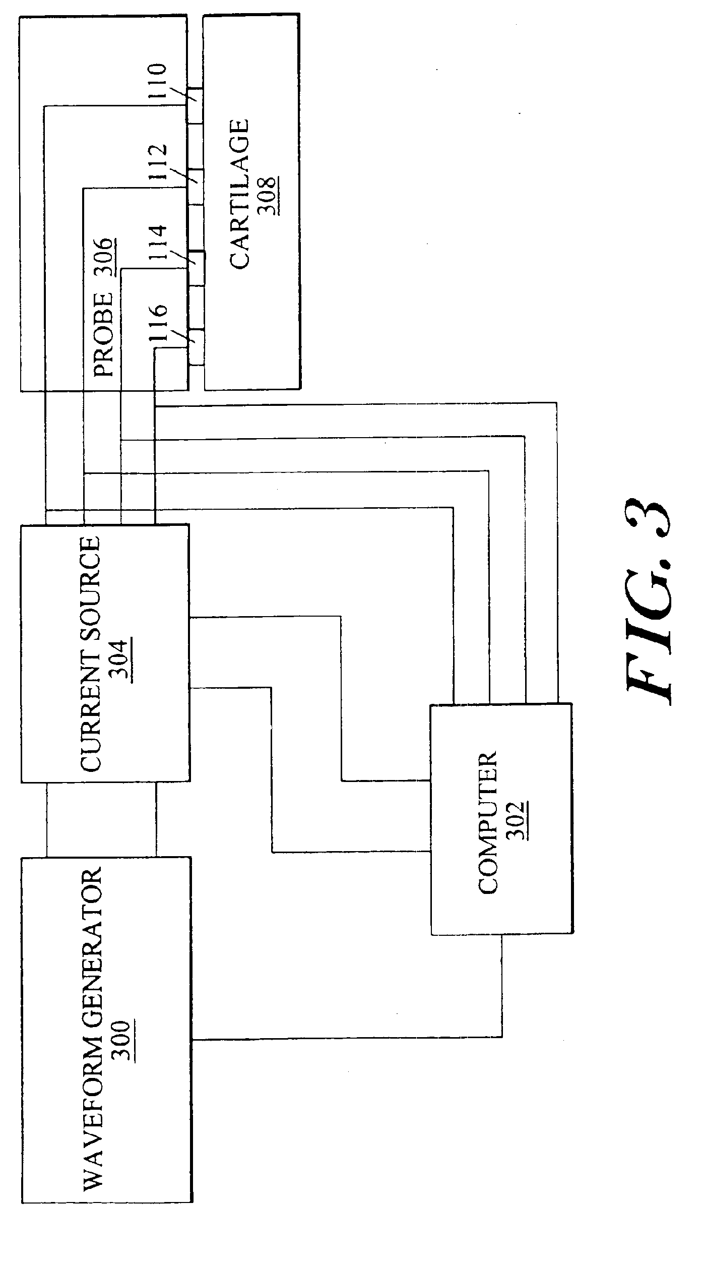
[0066]When current is applied to articular cartilage there is a distribution of current density and an electric field within the tissue, and also an associated voltage drop across the electrodes applying the current. The amplitude of the measured voltage drop between electrodes divided by the applied current amplitude is defined as the electrical impedance. The electrical impedance of the tissue for frequency ranges at least up to approximately 1 kHz, and possibly further, is dominated by the resistance of the hydrated ECM and this resistance, is inversely proportional to the density of mobile ions within the intratissue fluid. Thus, the electrical impedance of the tissue will increase with decreasing PG fixed charge content or increased swelling at constant PG content, since both these conditions lead to a lower concentration of mobile ions in the ECM by Donnan equilibrium. In addition, collagen network degradation will alter impedance.

[0067]This change in...


PUM


Abstract

The change in tissue impedance due to the change in the extracellular matrix that results from the degradation of cartilage is utilized to detect degradation of articular cartilage. A probe includes electrodes that apply a current to the articular cartilage which results in a current distribution and electric field within the cartilage, along with an associated voltage drop across the electrodes. The amplitude of this voltage drop is then measured and divided by the current applied to determine the tissue impedance. By measuring the impedance of patient tissue and comparing the detected patient impedance to a normal value for the tissue from clinically normal tissue, a determination of whether the patient tissue is degraded, and a determination of the extent of degradation is possible. Preferably, the impedance is measured using a probe with interdigitated electrodes. By changing which electrodes are utilized, the wavelength of the current distribution changes, allowing the probe to image depth dependent focal lesions.

Description

PRIORITY INFORMATION[0001]This application is a divisional application of Ser. No. 09 / 776,254 filed Feb. 2, 2001, now U.S. Pat. No. 6,735,468, which claims priority from provisional application Ser. No. 60 / 179,820 filed Feb. 2, 2000.[0002]This invention was made with government support under Subcontract No. AR 42285, under Prime Grant No. 2 R44 AR42285 02A1, awarded by the National Institutes of Health. The government has certain rights in the invention.BACKGROUND OF THE INVENTION[0003]The invention relates to the field of non-destructive arthroscopic diagnostic probes, and in particular to non-destructive arthroscopic diagnostic probes for detecting degeneration of articular cartilage utilizing impedance measurements.Articular Cartilage[0004]The function of organs in the human body are a direct consequence of their inherent structure. The function of an organ as a whole is more than the sum total of its individual constituents. Articular cartilage (AC) is a rich and illustrative ex...

Claims


Application Information

Patent Timeline
Patent Type & Authority Patents(United States)
IPC IPC(8): A61B5/103A61B5/05A61B5/053
CPCA61B5/05A61B5/053A61B5/103A61B5/4514A61B2562/0261A61B5/4528A61B5/6847A61B2562/0209A61B2562/0215
Inventor TREPPO, STEVENGRODZINSKY, ALAN J.QUAN, EMERSONFRANK, ELIOTBOMBARD, DAVIDBRESLAU, DAVID
Owner MASSACHUSETTS INST OF TECH
Why Eureka
Try Eureka