Method, system and computer readable medium for the two-dimensional and three-dimensional detection of lesions in computed tomography scans

a computed tomography and two-dimensional imaging technology, applied in tomography, image enhancement, instruments, etc., can solve the problems of fatigue or distraction, confusion in the identification of small lung nodules, and the prominence of blood vessels in ct images, so as to facilitate earlier diagnosis of lung cancer

Inactive Publication Date: 2005-05-24
ARCH DEVMENT
View PDF16 Cites 117 Cited by
  • Summary
  • Abstract
  • Description
  • Claims
  • Application Information

AI Technical Summary

Benefits of technology

[0031]The automated scheme for the detection of pulmonary nodules according to the present invention, advantageously, could provide substantial benefit in a screening program in which most cases will not contain nodules. Consequently, a computerized detection scheme, according to the present invention, implemented as a “second reader,” advantageously, is expected to help radiologists focus their attention on cases with regions that are suspicious for lung cancer. Advantageously, such a scheme could also be used to direct radiologists to suspicious lesions that would merit immediate targeted high-resolution scans following detection on the initial low-dose scan. Thus, the automated detection scheme according to the present invention could become an integral component of future lung cancer screening protocols.
[0032]The present invention has significance in that: (1) CT scans are used increasingly for diagnostic evaluation of patients, and (2) lung cancer screening by low-dose helical CT is gradually gaining acceptance as a viable means of early lung cancer detection. However, the large amount of image data acquired during a single CT examination may quickly lead to “information overload” for the radiologists who must interpret this data. The recent development of multi-slice CT acquisition will soon further compound the image information problem. Consequently, the automated lung nodule detection method according to the present invention is believed to be a valuable aid to radiologists, thereby facilitating earlier diagnosis of lung cancer.

Problems solved by technology

Although the potential camouflaging effect of overlapping anatomic structures is effectively eliminated in CT scans, identification of small lung nodules is still confounded by the prominence of blood vessels in CT images.
This process could lead to fatigue or distraction, especially when other abnormalities are present [10].

Method used


Image

  • Method, system and computer readable medium for the two-dimensional and three-dimensional detection of lesions in computed tomography scans
  • Method, system and computer readable medium for the two-dimensional and three-dimensional detection of lesions in computed tomography scans
  • Method, system and computer readable medium for the two-dimensional and three-dimensional detection of lesions in computed tomography scans

Examples


Embodiment Construction

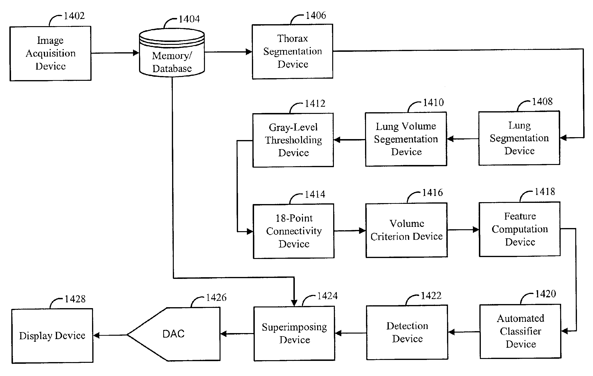
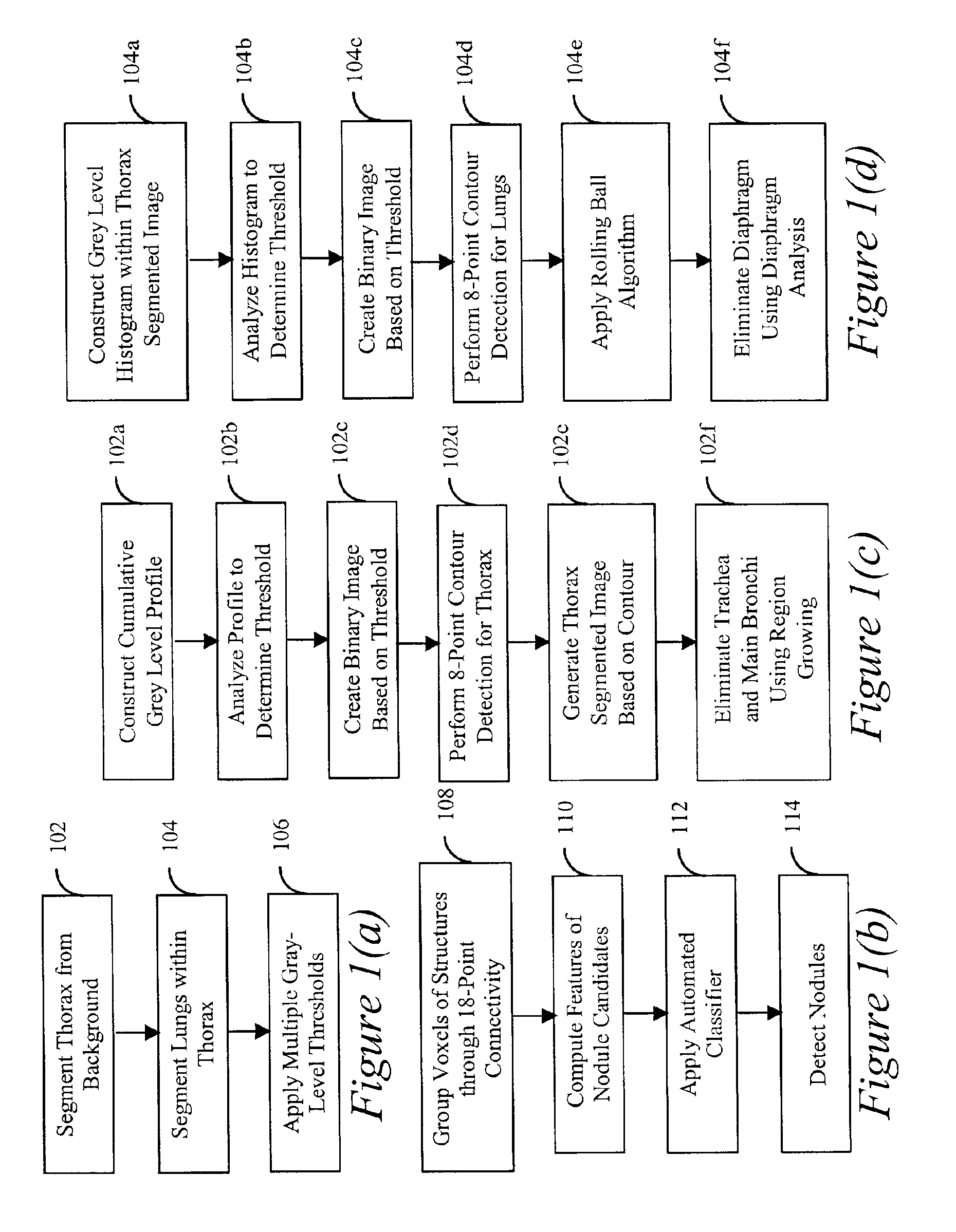
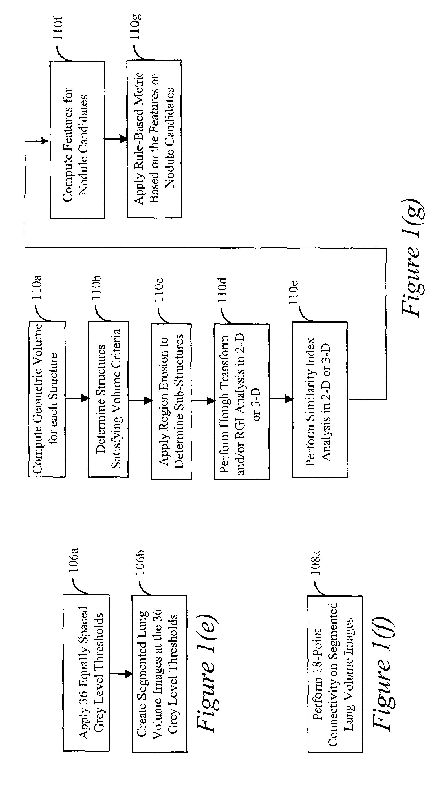
[0049]Referring now to the drawings, wherein like reference numerals designate identical or corresponding parts throughout the several views, and more particularly to FIGS. 1(a) and 1(b) thereof, there is illustrated a top-level block diagram of an automated method for the detection of lung nodules in thoracic CT scans according to the present invention, as is further discussed.

[0050]The overall scheme according to the present invention will now generally be described with reference to FIGS. 1(a) and 1(b) and will be later described in detail with reference to FIGS. 1(a)-(h), 2(a)-(b), 3, 4(a)-(b), 5(a)-(b), 6, 7(a)-(b), 8(a)-(d), 9-11, 12(a)-(b) and 13-15.

[0051]In FIGS. 1(a) and 1(b), the method according to the present invention includes initial acquisition of CT image data (not shown). At step 102, for each section image, a gray-level threshold is applied to create a binary image. A contour-detection algorithm is used to identify the outer margin of the largest “on” region in the...


PUM


Abstract

A method, system and computer readable medium for automated detection of lung nodules in computed tomography (CT) image scans, including generating two-dimensional segmented lung images by segmenting a plurality of two-dimensional CT image sections derived from the CT image scans; generating three-dimensional segmented lung volume images by combining the two-dimensional segmented lung images; determining three-dimensional lung nodule candidates from the three-dimensional segmented lung volume images, including, identifying structures within the three-dimensional segmented lung volume images that meet a volume criterion; deriving features from the lung nodule candidates; and detecting lung nodules by analyzing the features to eliminate false-positive nodule candidates from the nodule candidates.

Description

CROSS-REFERENCE TO RELATED APPLICATIONS[0001]This application claims priority under 35 U.S.C. §119(e) to U.S. provisional patent application Ser. No. 60 / 176,304, filed on Jan. 18, 2000, the entire contents of which is incorporated by reference herein.STATEMENT REGARDING FEDERALLY SPONSORED RESEARCH[0002]The present invention was made in part with U.S. Government support under USPHS grant numbers CA48985, CA62625, CA64370, and RR11459 (National Institute of Health). The U.S. Government has certain rights in the invention.BACKGROUND OF THE INVENTION[0003]1. Field of the Invention[0004]The present invention relates generally to a computerized method and system to aid radiologists in detection of abnormalities in tomography scans.[0005]The present invention also generally relates to computerized techniques for automated analysis of digital images, for example, as disclosed in one or more of U.S. Pat. Nos. 4,839,807; 4,841,555; 4,851,984; 4,875,165; 4,907,156; 4,918,534; 5,072,384; 5,133...

Claims


Application Information

Patent Timeline
Patent Type & Authority Patents(United States)
IPC IPC(8): G06T7/00A61B6/03G06T1/00G06T5/00G06T7/60G06T19/00
CPCG06T7/0012G06T7/0081G06T7/0097G06T2207/30064G06T2207/10124G06T2207/20061G06T2207/20141G06T2207/10081G06T7/11G06T7/174G06T7/187
Inventor ARMATO, III, SAMUEL G.GIGER, MARYELLEN L.MACMAHON, HEBER
Owner ARCH DEVMENT
Why Eureka
Try Eureka