
Processing EEG signals to predict brain damage

a brain damage and eeg technology, applied in diagnostic recording/measuring, instruments, applications, etc., can solve the problems of periventricular lesions, affecting the long-term neurological outcome, and being significantly less reliable in ultrasound imaging than mri

Inactive Publication Date: 2005-08-16
NATUS MEDICAL
View PDF2 Cites 96 Cited by
  • Summary
  • Abstract
  • Description
  • Claims
  • Application Information

AI Technical Summary

Benefits of technology

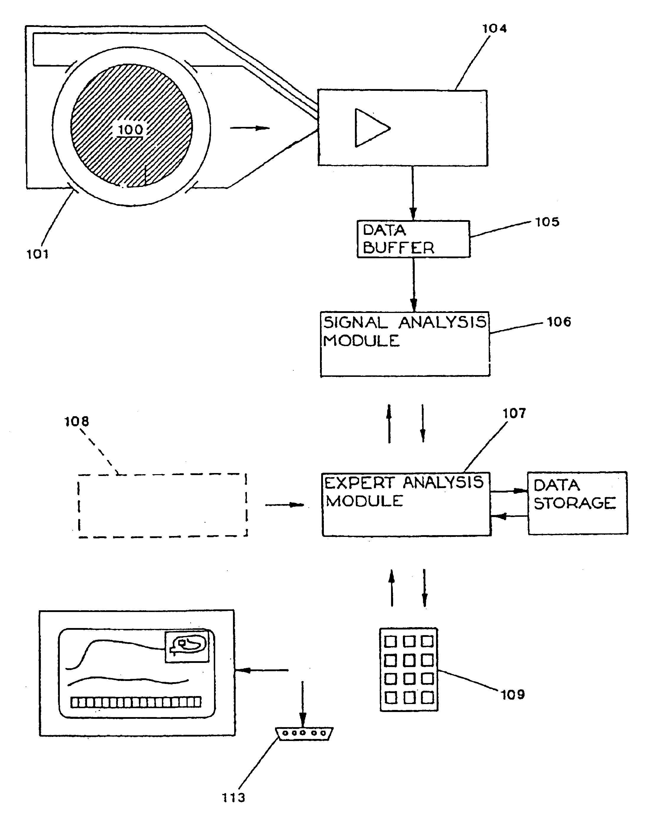
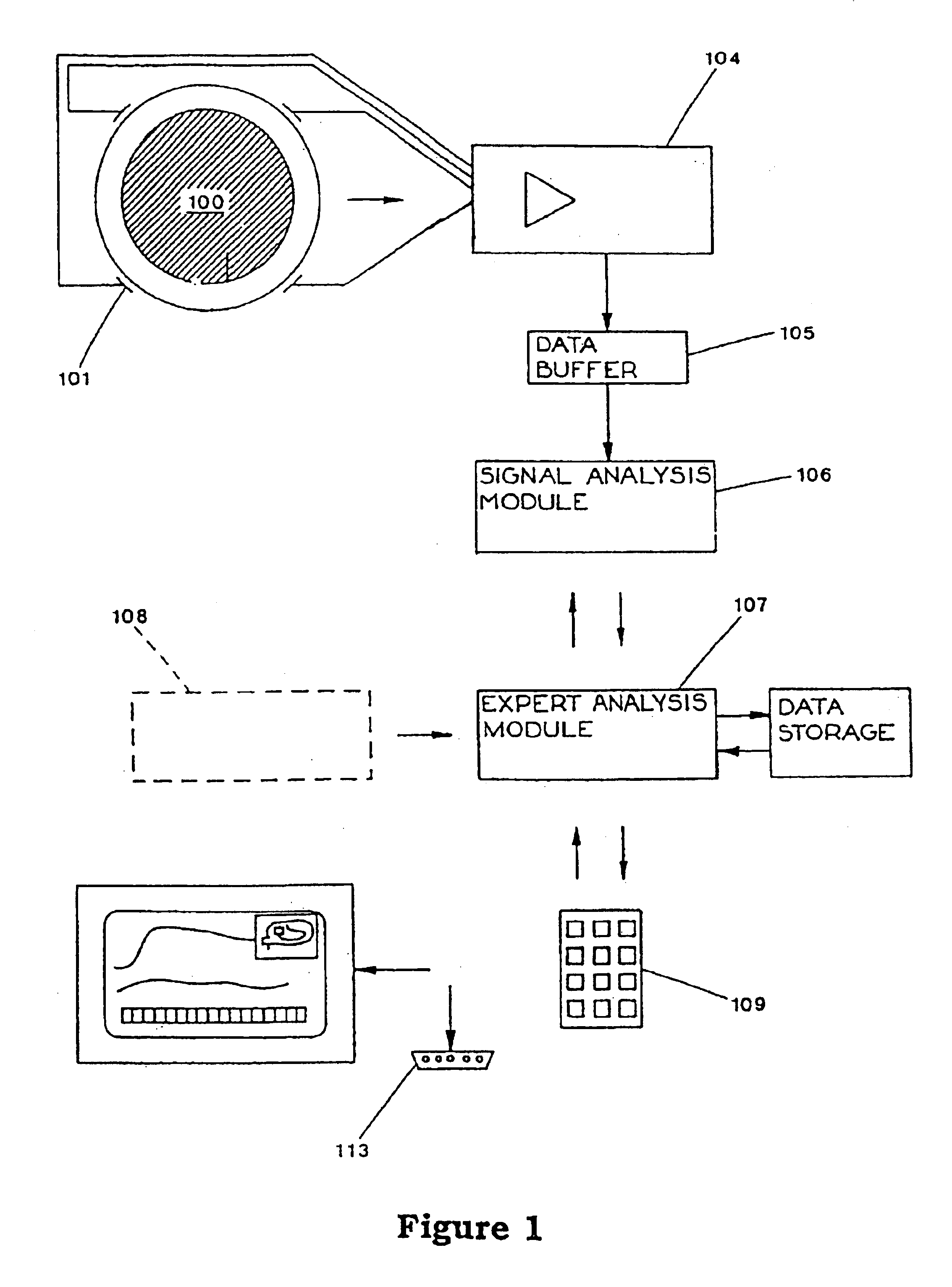
The invention is a device and method for detecting brain damage in newborns and predicting how well they will do. It uses an EEG machine to collect and analyze signals from the baby's brain to determine if there is any damage to the white matter, which is important for brain development. This information can help doctors manage the baby's care and predict how well they will do in life. The device is a computer software program that runs on a computer and collects and analyzes the EEG signals.

Problems solved by technology

Ultrasound imaging is significantly less reliable than MRI in this area.
Long term neurological outcome appears to be similarly compromised.
Also, both increased levels of hypoxanthine and prolonged metabolic acidosis in the neonatal period are associated with a high risk of periventricular lesions [Russel et al., Arch. Dis. Child., 67:388-392, 1992; Low et al., op. cit.].
Current methods for assessing brain injury reveal damaged areas of the brain, but do not identify those premature infants at risk of suffering a neural injury.
Brain damage assessed by neurological examination is of limited prognostic value, especially for those pre-term infants on life support.
However, this approach is less suitable for monitoring and detecting pathophysiologic events which may occur over several days, the knowledge of which could be used to minimize or avoid further injury.
However in surviving infants the timing of injuries is typically unclear and there are considerable problems with detecting when these white matter injuries occur [Murphy et al., Arch. Dis. Child Fetal Neonat. Ed., 75(1 Special Issue SI):F 27-F32, 1996].
The inability to detect the onset of injury makes management difficult.
Related to long-term EEG-based recordings, a further problem remaining to be solved is to reliably and consistently recover, over an extended period of perhaps 6 hours to a week, and in a non-ideal space, the very weak signal that comprises a typical EEG.
This signal is easily corrupted by intrinsic noise and external interference.

Method used


Image

  • Processing EEG signals to predict brain damage
  • Processing EEG signals to predict brain damage
  • Processing EEG signals to predict brain damage

Examples


Embodiment Construction

Definitions

[0044]An “electroencephalogram” or “EEG” comprises an outward indication of electrical activity from the most superficial layers of the cerebral cortex, usually recorded from electrodes on the scalp. This activity is the result of the rhythmic discharging of neurons under the electrode. The EEG signal provides information about the frequency and amplitude of the neuronal electrical activity and its temporal variation.

[0045]The “spectral edge frequency” is the xth percentile frequency of the EEG intensity over the frequency band 2 Hz to 20 Hz, i.e. the frequency below which lies x % of the total EEG intensity. Unless another percentile is specifically referred to, the “spectral edge frequency” is the 90th percentile frequency (more correctly “upper decile cortical spectral edge frequency”), and is the 90th percentile frequency of the EEG intensity over the frequency band 2 Hz to 20 Hz, i.e. the frequency below which lies 90% of the total EEG intensity.

[0046]“EEG intensity”...


PUM


Abstract

Rapid and accurate in-vivo assessment of cerebral white matter injury particularly for pre-term infants, for timely treatment and / or prediction of outcomes has been very limited. This invention exploits the discovery that reduced high-frequency EEG intensity, particularly as shown by the upper spectral edge frequency, is a good indicator of cerebral white matter neural injury and is well correlated with MRI results. With more experience of clinical cases, a set of simple rules such as “if the spectral edge value is below 8 Hz there is a high likelihood of injury” may be validated, yet the EEG technology involved is largely invisible to the user. In the invention, EEG signals are processed by software to obtain, store, and graphically display bilaterally collected EEG spectral edge and intensity values over from hours to weeks. Rejection of corrupted signals by filtering and gating means is responsive to incoming signal characteristics, to additional inputs such as motion sensors or impedance tests, and to patient data (gestational age in particular). The invention includes the software and methods of use.

Description

CROSS REFERENCE TO RELATED APPLICATIONS[0001]This application is a continuation-in-part of application Ser. No. 09 / 509,186, filed Apr. 7, 2000, now U.S. Pat. No. 6,493,577, application Ser. No. 09 / 509,196 is a 371 of PCT International Application No. PCT / NZ98 / 00142, filed Sep. 23, 1998, which in turn claims the priority of New Zealand Application No. 328820, filed Sep. 23, 1997. Each of these applications is incorporated into this application by reference.BACKGROUND OF THE INVENTION[0002]1. Field of the Invention[0003]This invention relates to a apparatus for display of a processed, condensed derivative of electroencephalographic (EEG) signals.[0004]2. Discussion of the Related Art[0005]Cerebral / neural injuries in pre-term infants are telatively difficult to assess and to track during their course, using skills and equipment available at the time of this application; yet intervention may be highly desirable. Magnetic resonance imaging (MRI) is the only reliable non-invasive assessme...

Claims


Application Information

Patent Timeline
Patent Type & Authority Patents(United States)
IPC IPC(8): G06F17/00A61B5/048A61B5/0476A61B5/374
CPCA61B5/048A61B5/4094A61B5/412A61B5/721A61B5/4806A61B5/7203A61B5/7257A61B5/726A61B5/7232A61B5/374
Inventor WILLIAMS, CHRISTOPHER EDWARD
Owner NATUS MEDICAL
Learn More