Opposed orthogonal fusion system and method for generating color segmented MRI voxel matrices

a color segmented, voxel-based technology, applied in the field of magnetic resonance imaging, can solve the problems of many limitations of gray tone methods in characterization, lack of automatic tissue segmentation that is required for accurate and immediate three-dimensional rendering of regions, and achieves the effect of being easy to understand by medical personnel

Inactive Publication Date: 2005-10-18
BROWN HUGH KEITH +1
View PDF10 Cites 37 Cited by
  • Summary
  • Abstract
  • Description
  • Claims
  • Application Information

AI Technical Summary

Benefits of technology

[0042]Another advantage of the invention is that a user studying MRI images can view a single color image that automatically characterized biophysical parameters for each tissue of interest, in contrast to multiple gray tone images representing each tissue.
[0043]Another advantage of the invention is that the several single color images can be combined for multidimensional viewing.
[0044]Another advantage is that the colorized three dimensional images obtained by the methods can disclose a combination of biophysical parameters typically undisclosed by a series of single parameter gray tone images.
[0045]Another advantage is that since the method of summative color composite tissue mapping is analogous to histologic staining, it is easily understood by medical personnel.

Problems solved by technology

Typically, gray tone methods have many limitations in the characterization since the human eye can typically only differentiate among 16 different gray tones.
However, even with successful production of a series of single composite images, there is typically still a problem of obtaining a segmented or characterized three dimensional rendering of the region of interest.
Even having several scans of several slices, there lacks a method of automatic tissue segmentation that is required for an accurate and immediate three dimensional rendering of regions of interest.

Method used


Image

  • Opposed orthogonal fusion system and method for generating color segmented MRI voxel matrices
  • Opposed orthogonal fusion system and method for generating color segmented MRI voxel matrices

Examples


Embodiment Construction

[0048]References to U.S. Pat. Nos. 5,332,968 and 5,410,250 are made throughout this description and are herein incorporated by reference.

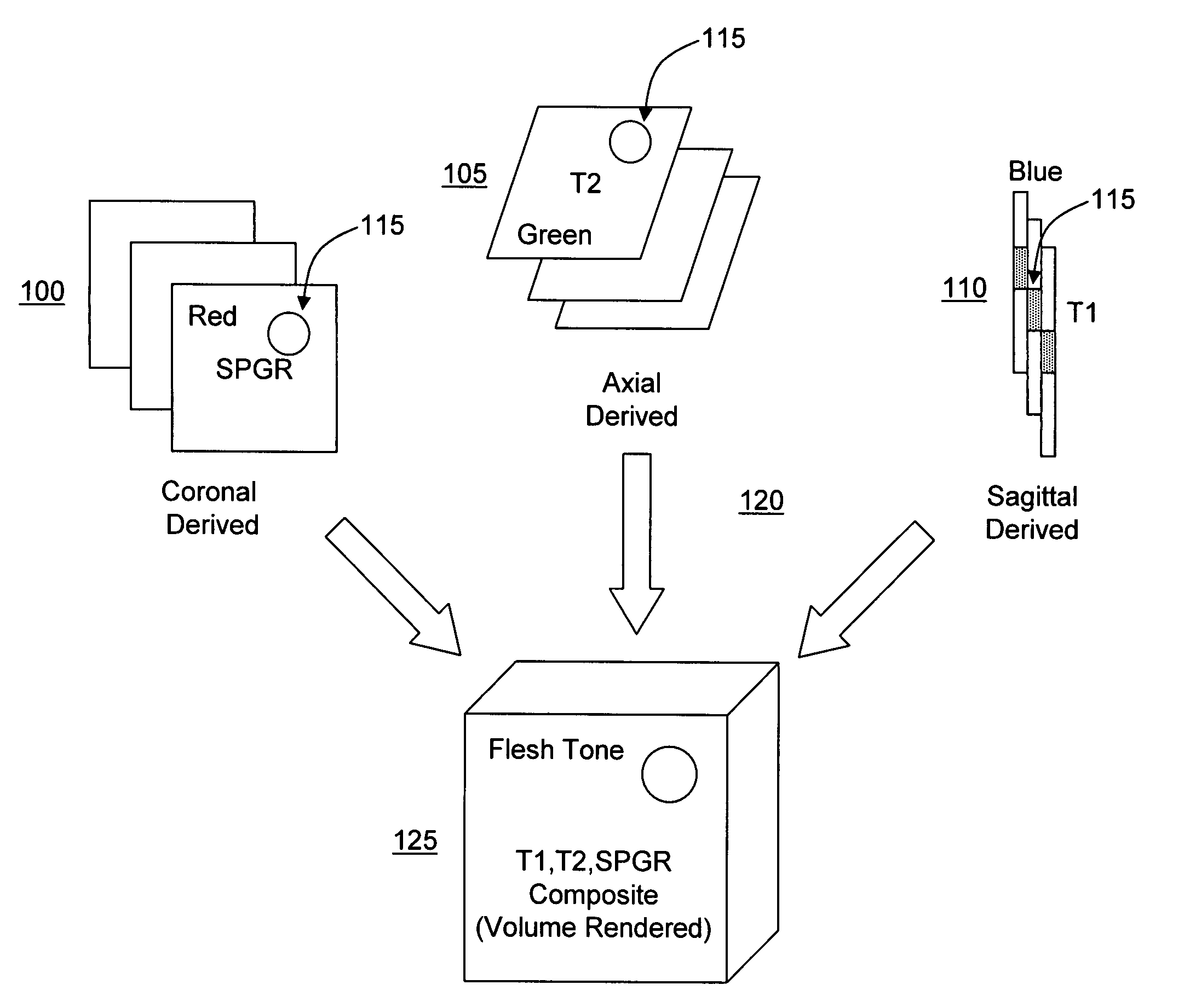
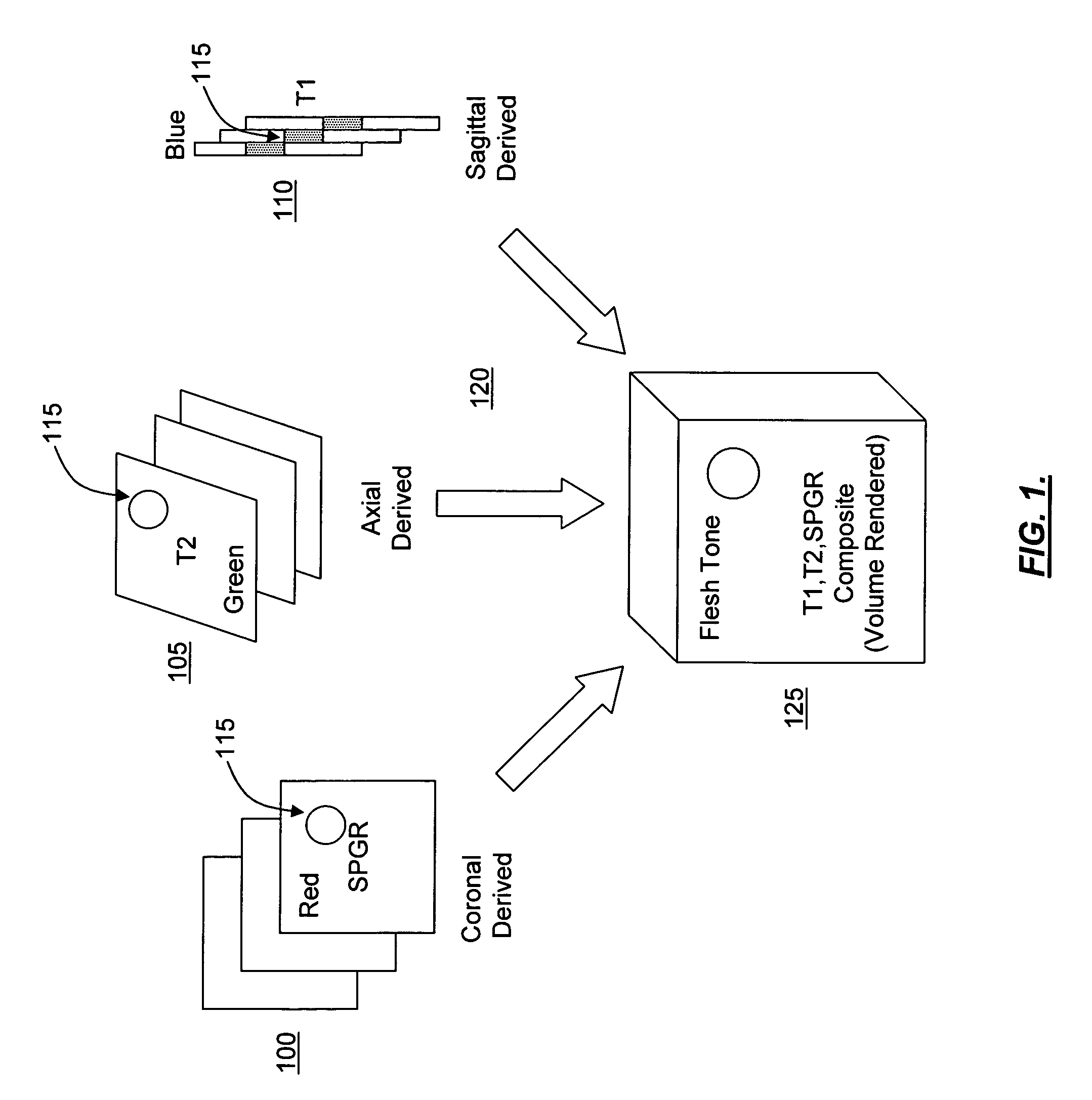
[0049]As achieved in earlier patents 5,332,968 and 5,410,250, automatic segmentation by assigning color to biophysical parameters of MRI images is made possible.

[0050]In general, this automatic segmentation is applied to segment, characterize, “tag”, or otherwise “label” different biophysical parameters of various tissue by unique color (RGB, HSI, CMY) is the prerequisite for 3D rendering without the various problems that exist today in characterizing images. In general, apparatuses and methods are described for producing a single color-coded composite image from a plurality of multi-parameter MRI image sets. Typically, these image sets are spatially aligned and are acquired using different pulse sequences to contrast various parameters of anatomical, physiological and pathological features. The methods include acquiring gray tone images and plotti...


PUM


Abstract

Systems and methods for producing multi-dimensional tissue characterization images are described. By combining colorized MRI images composited from multiple gray tone MRI images, riffle stacks can be formed and combined to render multidimensional reconstruction of anatomical, physiological and pathological features. Biophysical parameters are therefore automatically segmented, characterized, tagged, mapped and otherwise labeled. In another embodiment, gray tone images are collected by acquiring orthogonally opposed pulse sequences, the images of which are color masked and uniquely rendered in three dimensions to obtain voxels in which the color is determined by the summative color masks applied to original gray tone pixels. This method can be embodied as specific applications designed for the brain, abdomen, breast, pelvis, musculoskeletal and other organs and biophysical systems to facilitate diagnosis, surgical planning and virtual exploration.

Description

[0001]This application is a utility application which claims the benefit of priority of U.S. Provisional Patent Application No. 60 / 345,393, filed on Jan. 2, 2002, entitled “MRI Image Processing”.BACKGROUND OF THE INVENTION[0002]I. Field of the Invention.[0003]The present invention relates generally to the field of magnetic resonance imaging (MRI) and more particularly to systems and methods for producing multi-dimensional tissue characterization images.[0004]II. Description of the Related Art[0005]MRIs are used for a variety of reasons such as medical diagnosis, surgical planning, and enhanced visualization of anatomic, physiologic and pathologic features. Radio frequency signals from three dimensional tissue voxels are absorbed and then radiated when the tissue is placed in a strong magnetic field. These signals provide data for the spatial location and contrast discrimination of specific tissues, which are ultimately displayed as pixels comprising a two-dimensional MR image. These...

Claims


Application Information

Patent Timeline
Patent Type & Authority Patents(United States)
IPC IPC(8): G01V3/00G06K9/00H04N9/00H04N9/43G01R33/56
CPCG01R33/56
Inventor BROWN, HUGH KEITHMOODY, JENNIFER D.
Owner BROWN HUGH KEITH
Why Eureka
Try Eureka