Methods and apparatus for securing medical instruments to desired locations in a patient's body

a technology for securing medical instruments and patient bodies, applied in applications, vaccination/ovulation diagnostics, surgery, etc., can solve the problems of reducing the likelihood of removing target tissue, patient's breast must remain, and no single procedure is ideal for all cases, so as to achieve accurate placement

Inactive Publication Date: 2005-10-25
SENORX
View PDF60 Cites 137 Cited by
  • Summary
  • Abstract
  • Description
  • Claims
  • Application Information

AI Technical Summary

Problems solved by technology

The type of biopsy utilized depends in large part on circumstances present with respect to the patient, including the location of the lesion(s) within the body, and no single procedure is ideal for all cases.
However, it often occurs that the barb or hook at the distal end of the wire marker attaches to something other than the tumor or lesion.
However, breast tissue is comprised of fibrous bands which, in compression, may be close to the target lesion and inadvertently engaged by the barb of the localization wire.
As in the case of localization wires, there is some risk in using any of the foregoing devices that the distal end of the instrument will migrate away from the target lesion during the biopsy procedure, thereby reducing the likelihood of removing target tissue.
One disadvantage of this approach, however, is that the patient's breast must remain in compression during the entire procedure, with attendant discomfort and increased procedural difficulty, in order to properly utilize the imaging equipment.
Furthermore, stereotactic imaging equipment or other suitable alternatives can cost as much as $400,000 or more and is not in the usual inventory of a typical community hospital.

Method used


Image

  • Methods and apparatus for securing medical instruments to desired locations in a patient's body
  • Methods and apparatus for securing medical instruments to desired locations in a patient's body
  • Methods and apparatus for securing medical instruments to desired locations in a patient's body

Examples


first embodiment

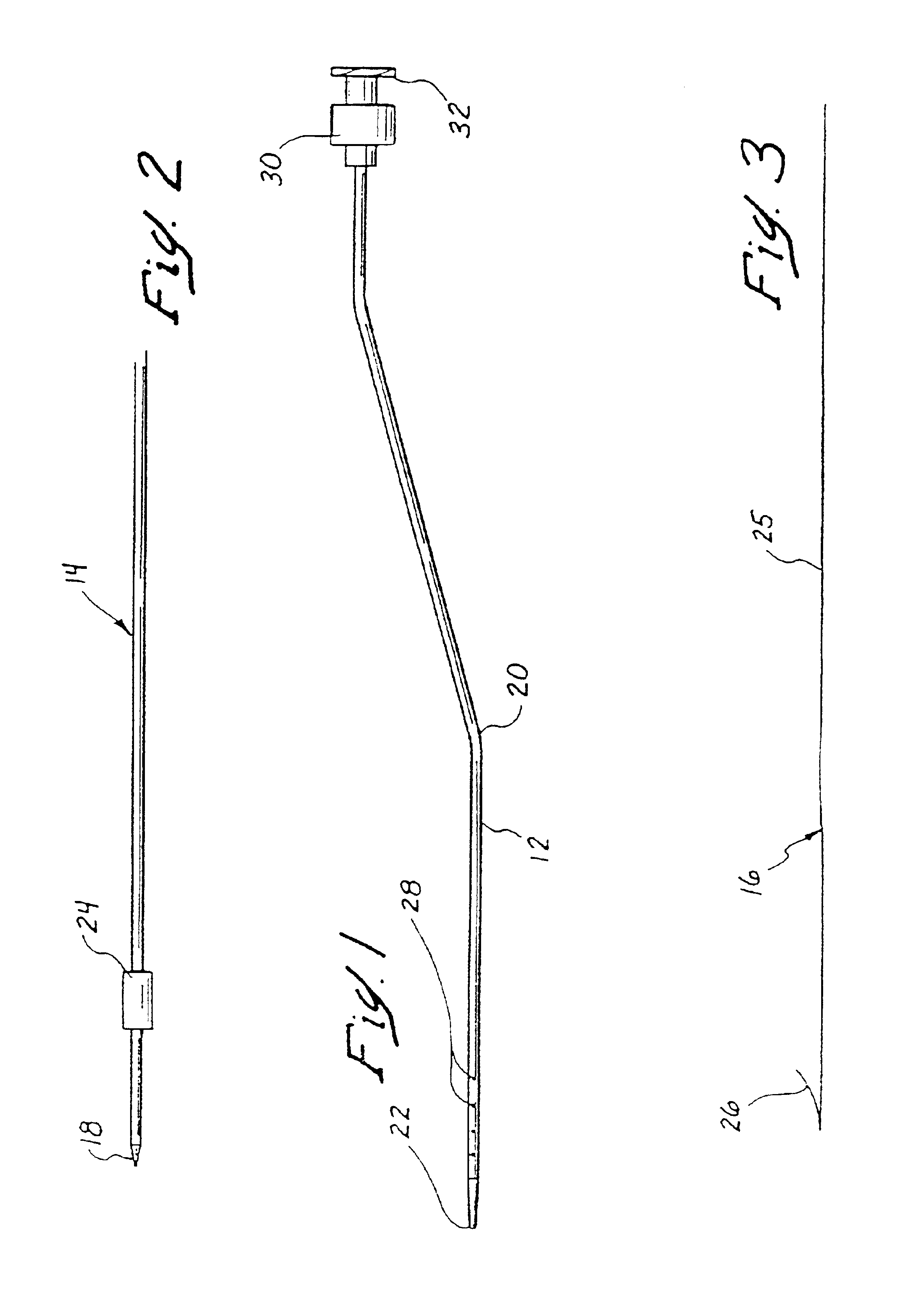
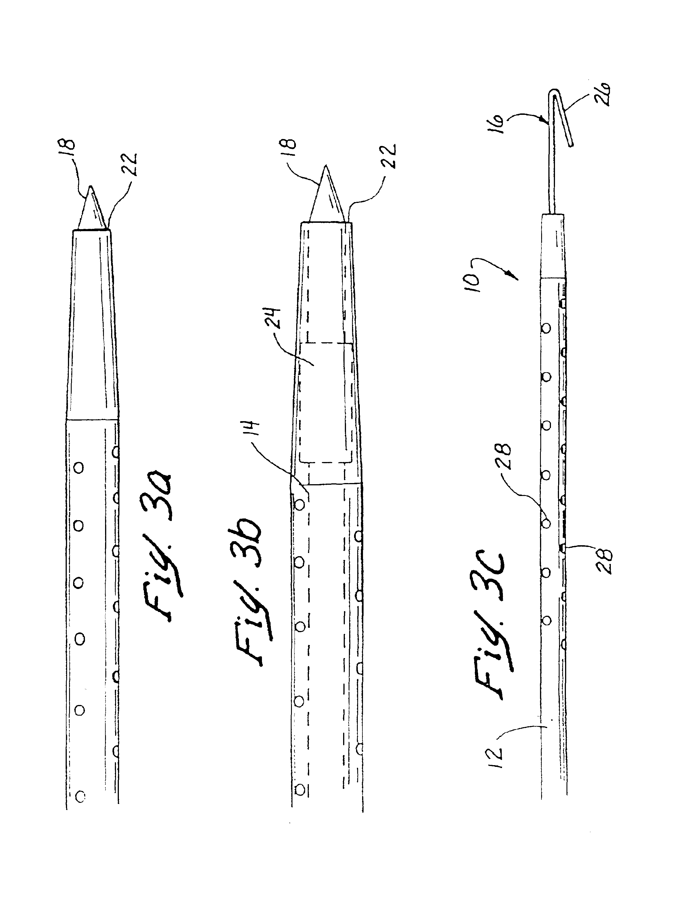
[0044]Referring now more particularly to the drawings, FIGS. 1-3c illustrate the invention, wherein a medical instrument 10 (FIGS. 3a-3c) comprises a catheter 12 (FIGS. 1, 3a-3c), an introducer needle 14 (FIGS. 2, 3a, and 3b), and a localization wire 16 (FIGS. 3, 3c). In this embodiment, which may be styled as a “needle in” infusion system, the introducer needle 14 comprises a sharp distal end 18, which is inserted through an entry hole 20 in the catheter 12 (FIG. 1), so that its tip 18 extends beyond the distal end 22 of the catheter 12, as shown in FIGS. 3a and 3b. The introducer needle 14 may include a stop 24 having an enlarged diameter, which is adapted to engage the distally tapering inner sidewall of the catheter 12 at a predetermined point, as generally shown particularly in FIG. 3b, to ensure that the tip 18 properly extends beyond the distal end 22 of the catheter 12. The introducer needle 14 and catheter 12 together are then introduced into a patient's body (not shown), u...

third embodiment

[0054]Still a third embodiment, which functions in a manner equivalent to that of a localization wire is illustrated in FIGS. 9 and 10. In this embodiment, a catheter 48, which comprises a proximal hub 50, a distal end 52, and a lumen 54, is insertable into a patient's body using conventional image guidance techniques, so that the distal end 52 is disposed at a desired target tissue site. Once properly located, a bonding agent 33 is infused through one or more infusion ports 56 to surrounding target tissue, in order to bond the distal end of the catheter 48 to the surrounding tissue. Again, as in the previous embodiments, the bonding agent may be injected into the lumen 54 of the catheter through the proximal hub 50, or may alternatively be stored in the distal end 52 of the catheter and selectively released at the desired time.

[0055]FIGS. 11 and 12 illustrate two alternative embodiments for the outer tube 58 of the catheter in any of the foregoing embodiments. In FIG. 11, the tube ...


PUM


Abstract

Devices and methods are provided for securely affixing a medical instrument to desired tissue in a patient's body, using a fixation agent. Such medical instruments may comprise localization wires or tissue acquisition instruments, such as biopsy instruments, for example. In the case of tissue acquisition instruments, the inventors have discovered significant advantages for securely affixing the distal end of the tissue acquisition instrument to a particular tissue target area. For example, such an approach permits the imaging environment to be uncoupled from the procedural environment so that expensive and often unavailable imaging equipment, such as stereotactic imaging equipment, need not be used. In a preferred embodiment, a bonding agent, such as adhesive, surgical glue, or a solvent, is used as the fixation agent.

Description

[0001]This application is a continuation of patent application Ser. No. 09 / 146,185, filed Sep. 1, 1998, now 6,540,693 which is a continuation-in-part of patent application Ser. No. 09 / 057,303, filed Apr. 8, 1998 now 6,331,166, which claims benefit to provisional patent application Ser. No. 60 / 076,973, filed Mar. 3, 1998, all of which applications are hereby incorporated herein by reference in their entirety and from which priority is hereby claimed under 35 U.S.C. §§119(e) and 120.FIELD OF THE INVENTION[0002]The present invention relates to methods and devices for ensuring that a medical instrument remains in a desired location within a patient's body during a medical procedure, and more particularly to methods and devices for affixing a distal end of the medical instrument to the desired location using adhesives.BACKGROUND OF THE INVENTION[0003]It is often desirable and frequently necessary to sample or remove a portion of tissue from humans and other animals, particularly in the d...

Claims


Application Information

Patent Timeline
Patent Type & Authority Patents(United States)
IPC IPC(8): A61B17/32A61B18/14A61B10/00A61B17/00A61B17/22A61B19/00A61B17/34A61B10/02A61B18/12
CPCA61B10/0241A61B10/0266A61B17/00491A61B17/32056A61B18/14A61B18/1477A61B19/54A61B10/02A61B17/320725A61B17/3417A61B18/1487A61B18/1492A61B19/5225A61B2017/320064A61B2017/3488A61B2018/00208A61B2018/00214A61B2018/00267A61B2018/00333A61B2018/00577A61B2018/00898A61B2018/0091A61B2018/00916A61B2018/1253A61B2018/126A61B2018/1407A61B2018/1425A61B2018/1475A61B2018/162A61B2019/205A61B2019/5408A61B90/17A61B90/37A61B90/39A61B2090/3908A61B2090/3987A61B10/04A61B2090/3991A61B18/12A61B2018/00601
Inventor BURBANK, FRED H.LUBOCK, PAULJONES, MICHAEL L.QUICK, RICHARD L.
Owner SENORX
Why Eureka
Try Eureka