

Tomographic microscope for high resolution imaging and method of analyzing specimens
a tomographic microscope and high-resolution technology, applied in the field of computerized tomographic microscopes for evaluating specimens, can solve the problems of affecting the quality of specimens, so as to achieve the effect of removing undesirable information
Inactive Publication Date: 2006-02-21
HEWITT CHARLES W +4
View PDF12 Cites 50 Cited by
- Summary
- Abstract
- Description
- Claims
- Application Information
AI Technical Summary
Benefits of technology
[0014]In a preferred embodiment of the method, in step (2), the information cross-correlated comprises absorbed or emitted light frequency information and intensity for each pixel. Most preferably, the cross-correlation comprises a cross correlation of information between a linearly arranged sequence of pixels from each image, the sequences at least partially overlapping. Furthermore it is preferred that the cross-correlation will, for purposes of constructing a confocal emulated image, score as undesirable that specific image information that is displaced in one image relative to the other and will score as desirable that specific image information that is not displaced in one image relative to the other, and further for purposes of constructing a confocal emulated image will tend to retain desirable information and to eliminate undesirable information.
[0024]It is preferred that the means for cross-correlation will, for purposes of constructing a confocal emulated image, score as undesirable that specific image information that is displaced in one image relative to the other and will score as desirable that specific image information that is not displaced in one image relative to the other, and further for purposes of constructing a confocal emulated image will tend to retain desirable information and to eliminate undesirable information.
Problems solved by technology
For example, the standard compound microscope using axial illumination is particularly ill-suited for clusters of cells.
Thick or overlapping images can be distorted because of diffraction of the light and because of absorption by thick sections.
Moreover, depth of focus is greatly reduced at high power, and it is difficult to visualize structures above or beyond a thin focal plane.
Evaluation is made difficult by deficiencies in resolution, contrast, light penetration, and sharpness of image.
As a consequence of the difficult and subjective nature of the test, there is a continuing problem with false negative readings and ambiguous readings in various cytopathology specimens.
Microscopic observation of thick cell clusters proved difficult as is well known to those skilled in the art using conventional axial illumination, as interpretation within the flat field is suboptimal.
To achieve this goal, however, a number of significant challenges must be met.
One challenge results from the nature of the image the computer “sees” and the relationship of that seen image to the actual image in the specimen under the microscope.
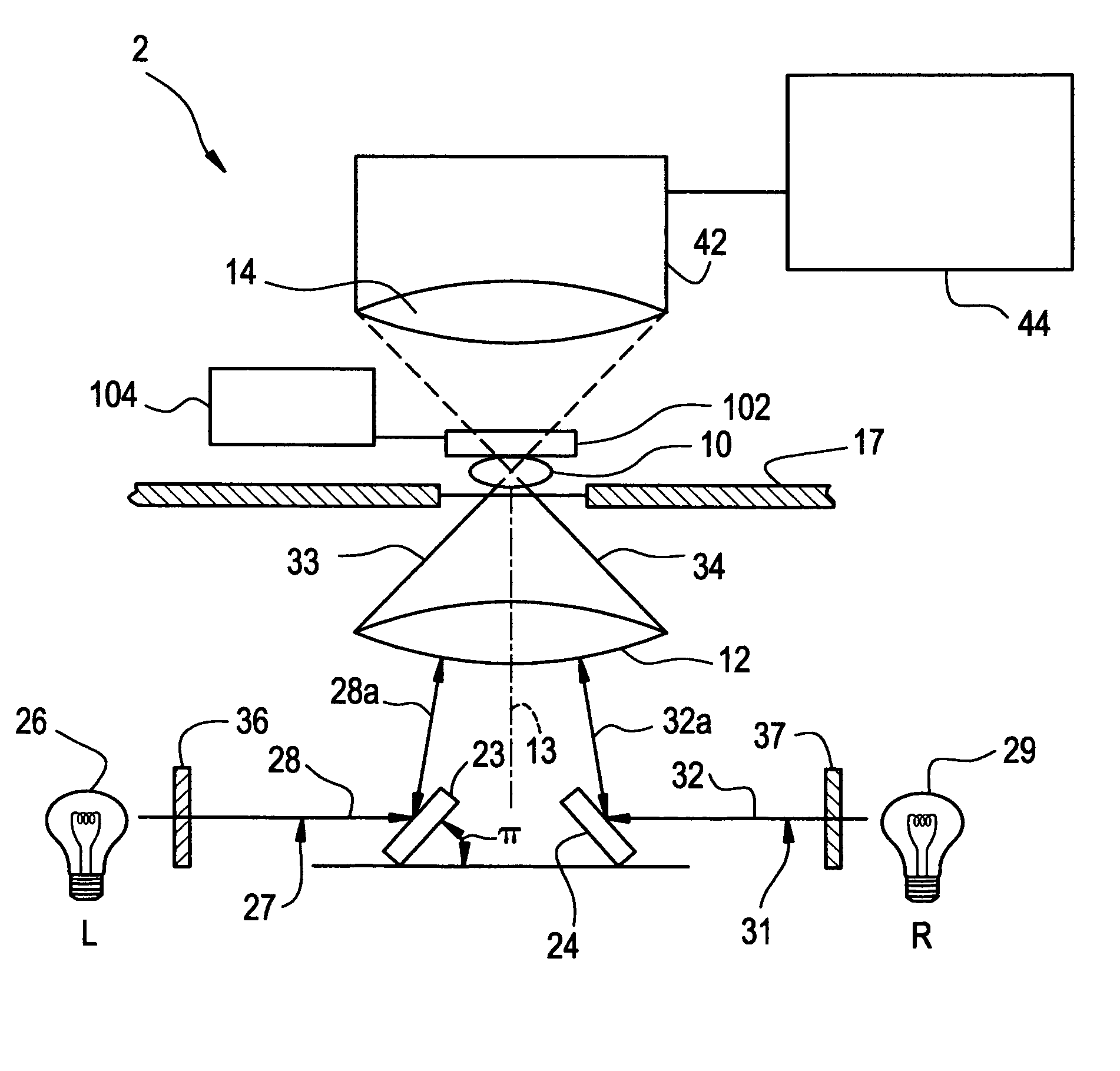
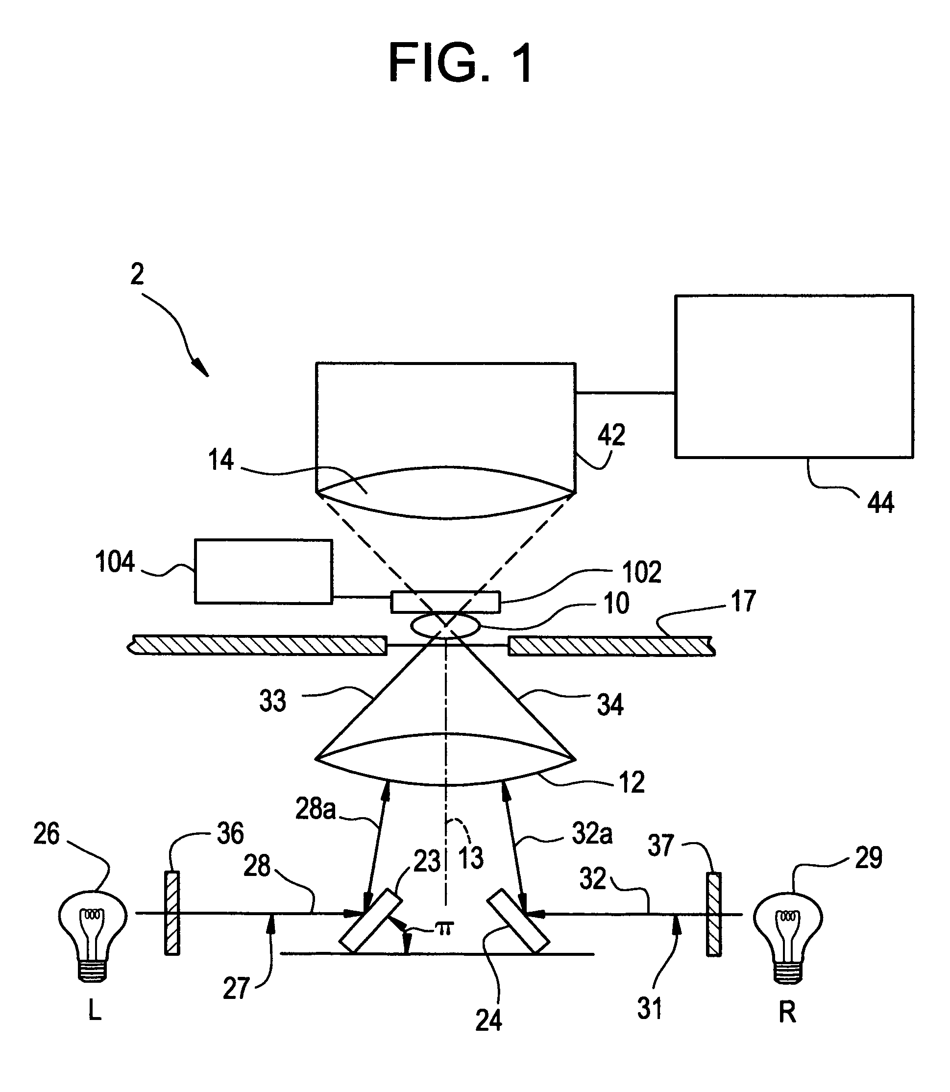
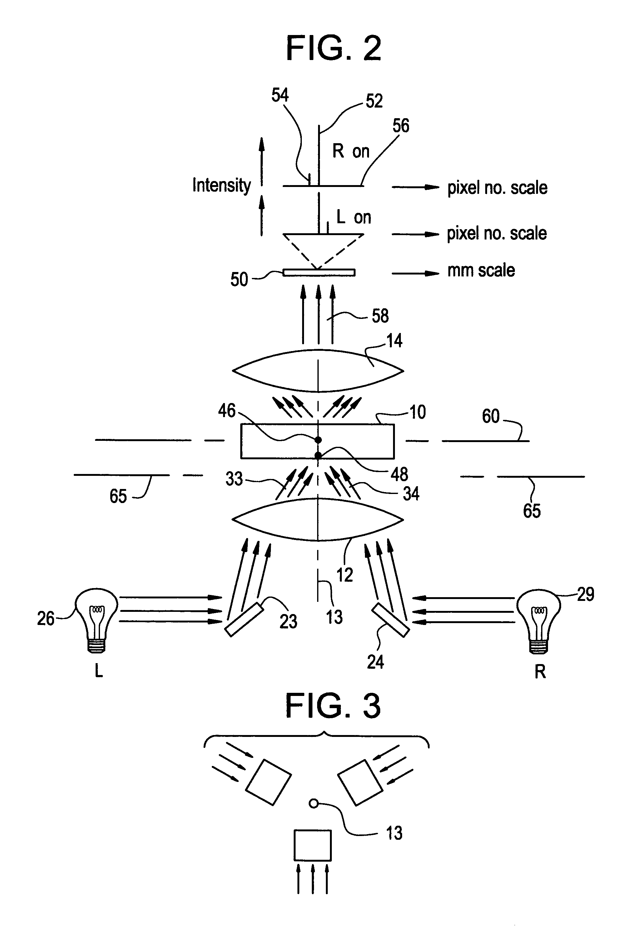
Because, however, microscopes cannot be perfectly focused, the pixels seen by the computer will have received light not only from the focal plane of interest (i.e., a “virtual” thin slice of the specimen), but also specimen components just below and just above that plane.
Method used
View moreImage
Smart ImageViewing Examples

Examples
examples
[0069]Optical sections of thick (10 to 40 micron) cell clusters from cervical Pap smears were captured sequentially at 400× using various oblique light paths. Images were then processed with a computer algorithm to produce confocal emulation. Three-dimensional reconstructions were then complied with the high-resolution images using Voxblast software (available from VayTek, Inc. of Fairfield, Iowa). Cells were color segmented, and various volumetric characteristics were emphasized.
[0070]An illustration of the result of using the method of the invention is shown in FIG. 5.
Login to View More PUM
 Abstract
Tomographic methods and device for obtaining images of microscopic specimens such as Pap smears.
Description
[0001]This application claims the benefit of U.S. provisional application Ser. No. 60 / 163,166 filed Nov. 2, 1999.FIELD OF THE INVENTION[0002]The present invention relates to a computerized tomographic microscope for evaluating specimens at high-resolution and a method of analyzing a specimen with use of a high-resolution three-dimensional reconstruction.BACKGROUND OF THE INVENTION[0003]The proper evaluation of pathological specimens is of high importance to public health. An example is the detection and diagnosis of cancer. Often this is performed by a screening mechanism in which a cell specimen is obtained from a patient and detected under a microscope for abnormalities. While screening tests have decreased mortalities associated with cancer by preventing the development of invasive disease, it is nonetheless dependent on the ability of a technician viewing a specimen under a microscope to detect abnormal cells and structures in thick specimens or thick clusters of cells. Success ...
Claims
Login to View More Application Information
Patent Timeline
 IPC IPC(8): G06K9/00
CPCG06K9/00134G06V20/693
InventorHEWITT, CHARLES W.DOOLIN, EDWARD J.KESTERSON, JOHNLAUREN, PETER D.GREENBERG, GARY
OwnerHEWITT CHARLES W