Method and system for lung disease detection

a lung disease and image data technology, applied in the field of medical image data processing, can solve the problems of affecting the sensitivity and specificity of human reading, time-consuming and labor-intensive radiologists, and difficult for radiologists to classify the extent of disease progression

Inactive Publication Date: 2006-06-06
GENERAL ELECTRIC CO
View PDF18 Cites 59 Cited by
  • Summary
  • Abstract
  • Description
  • Claims
  • Application Information

AI Technical Summary

Problems solved by technology

The considerable volume of data presented by a single CT scan presents a time-consuming process for radiologists.
Fatigue is therefore a significant factor affecting sensitivity and specificity of the human reading.
In other diseases, such as emphysema, it is difficult for a radiologist to classify the extent of disease progression by only looking at the CT images.
The above techniques presented to date, however, have largely focused on identifying suspicious lesions in CT scans and have not directly addressed obtaining correct differentiation of structures in the lung and correct measurements of their size.
Additionally, the above techniques are generally limited in the interpretative nature of the results.
There has been no apparent evaluation by the above techniques to address the type of or characteristics of the scanning device in the analysis of the images produced.

Method used


Image

  • Method and system for lung disease detection
  • Method and system for lung disease detection
  • Method and system for lung disease detection

Examples


Embodiment Construction

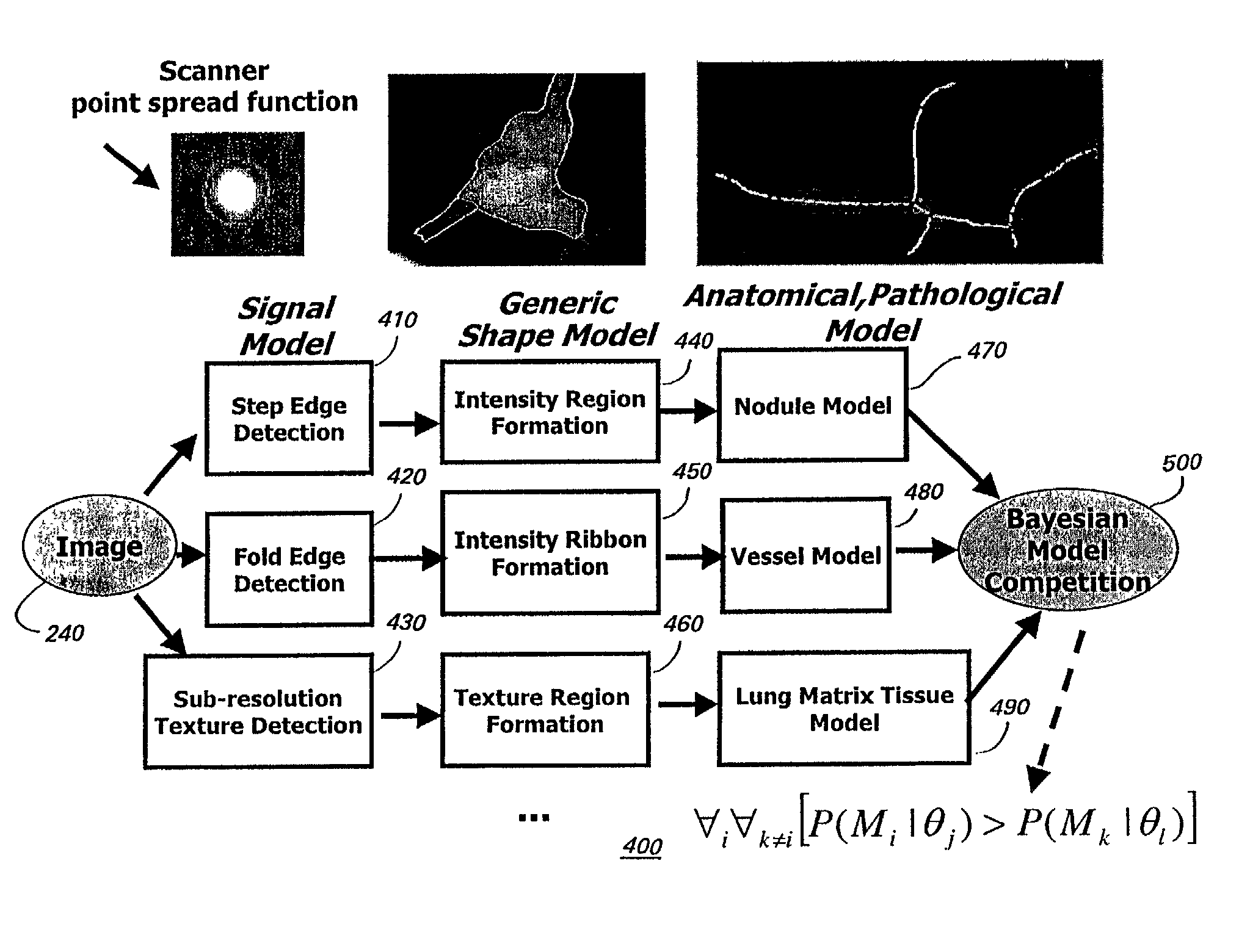
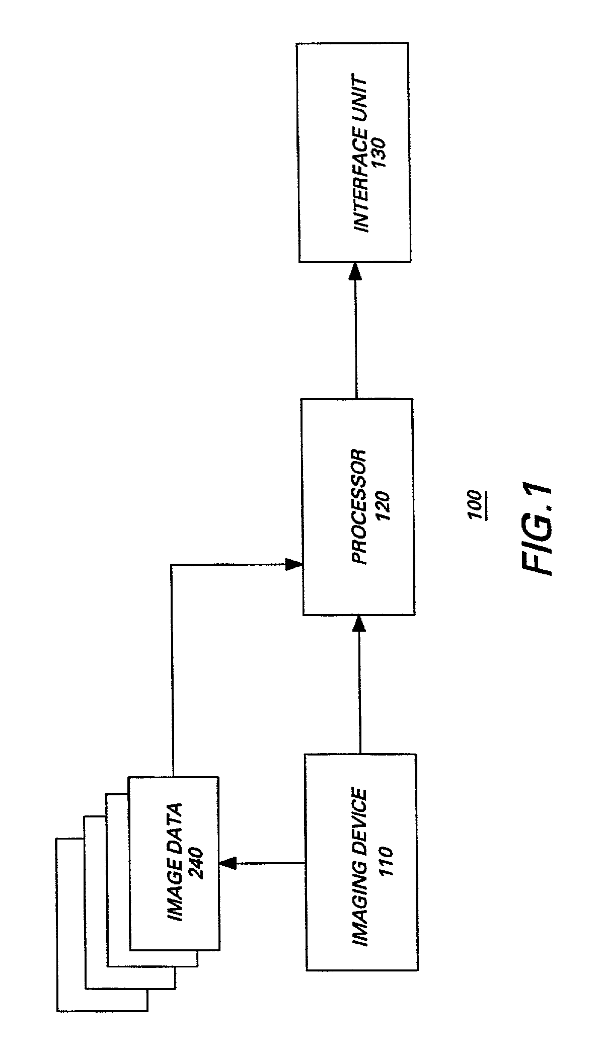
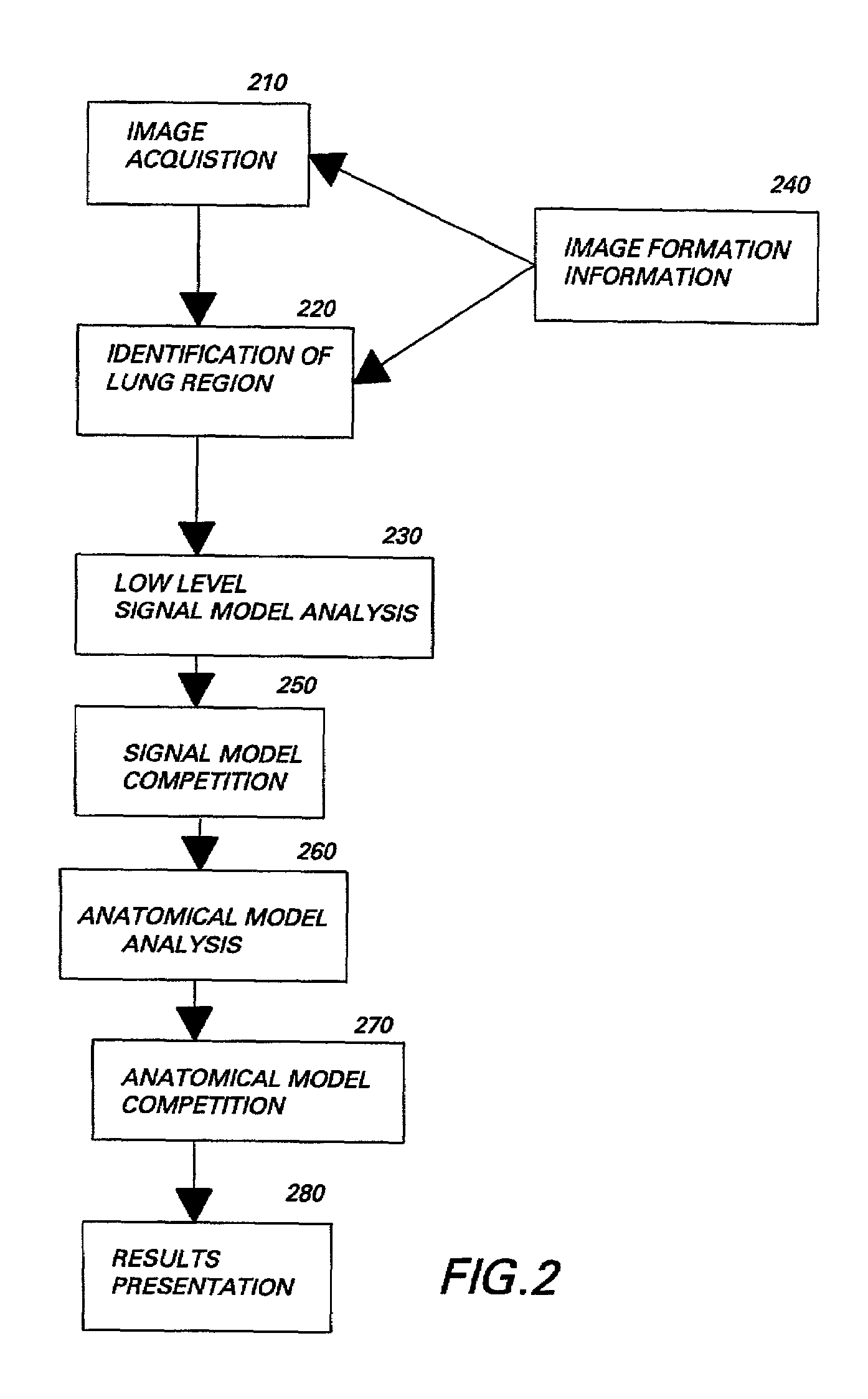
[0016]Referring to FIG. 1, a general block diagram of a system 100 for disease detection is shown. System 100 includes an imaging device 110, which can be selected from a number of medical imaging devices known in the art for generating a plurality of images. Most commonly, computed tomography (CT) and magnetic resonance imaging (MRI) systems are used to generate a plurality of medical images.

[0017]During a CT imaging session, a patient lies horizontal and is exposed to a plurality of x-rays measured with a series of X-ray detectors. A beam of x-rays passes through a particular thin cross-section or “slice” of the patient. The detectors measure the amount of transmitted radiation. This information is used to compute the x-ray attention coefficient for sample points in the body. A gray scale image is then constructed based upon the calculated x-ray attenuation coefficients. The shades of gray in the image contrast the amount of x-ray absorption of every point within the slice. The sl...


PUM


Abstract

A method for processing medical images for use in the detection and diagnosis of disease comprises classifying regions of interest within the medical images based on a hierarchy of anatomical models and signal models of signal information of an image acquisition device used to acquire the medical images. The anatomical models are derived to be representative of anatomical information indicative of a given disease. A computer-aided system for use in the diagnosis and detection of disease comprises an image acquisition device for acquiring a plurality of image data sets and a processor adapted to process the image data sets. The processor is adapted to classify selected tissue types within the image data sets based on a hierarchy of signal and anatomical models and the processor is further adapted to differentiate anatomical context of the classified tissue types for use in the diagnosis and detection of disease.

Description

BACKGROUND OF INVENTION[0001]This invention relates to a method and system for processing medical image data to aid in the detection and diagnosis of disease, and more particularly, to a method and system for detecting lung disease in medical images obtained from a x-ray computed tomography (CT) system.[0002]A x-ray chest radiograph system is the more commonly used diagnostic tool useful for the purpose of detecting lung disease in humans. Lung disease such as bronchitis, emphesema and lung cancer are also detectable in chest radiographs and CT. However, CT systems generally provide over 80 separate images for a single CT scan thereby providing a considerable amount of information to a radiologist for use in interpreting the images and detecting suspect regions that may indicate disease.[0003]Suspect regions are defined as those regions a trained radiologist would recommend following through subsequent diagnostic imaging, biopsy, functional lung testing, or other methods. The consid...

Claims


Application Information

Patent Timeline
Patent Type & Authority Patents(United States)
IPC IPC(8): G06K9/00G01R33/54A61B5/055A61B6/00A61B6/03A61B8/00G01T1/161G06T7/00
CPCA61B6/00G06T7/0012G06T2207/30061
Inventor MUNDY, JOSEPH LEAGRANDMCCULLOCH, COLIN CRAIGAVILA, RICARDO SCOTTHASTINGS, SHANNON LEEKAUCIC, JR., ROBERT AUGUSTLORENSEN, WILLIAM EDWARDTUREK, MATTHEW WILLIAM
Owner GENERAL ELECTRIC CO
Why Eureka
Try Eureka