Variational approach for the segmentation of the left ventricle in MR cardiac images

a technology of cardiac images and variables, applied in the field of system and method for segmenting cardiac images, can solve the problems of model sensitive to the initial conditions, physiology, cardiology, and cannot fully exploit the prior knowledge available in other domains, and achieve the effect of avoiding noise or physical corruption, avoiding the occurrence of abnormalities, and avoiding abnormalities

Inactive Publication Date: 2006-07-18
SIEMENS CORP
View PDF4 Cites 61 Cited by
  • Summary
  • Abstract
  • Description
  • Claims
  • Application Information

AI Technical Summary

Benefits of technology

[0018]In one aspect of the invention, a method for segmenting a cardiac image comprises receiving a cardiac image, determining boundary information for one or more contours in the cardiac image using a gradient vector flow boundary method and determining region information to separate regions in the cardiac image

Problems solved by technology

However, these techniques fail to take fully advantage of the available prior knowledge from other domains such as physiology, cardiology, etc.
Although this primitive approach has led to outstanding results, it can be sensitive to noisy or physically corrupted data.
However, these models are quite sensitive to the initial conditions.
This condition, however, cannot be easily met for general medical image segmentation applications.
Although various techniques have been proposed to deal with these issues, th

Method used


Image

  • Variational approach for the segmentation of the left ventricle in MR cardiac images
  • Variational approach for the segmentation of the left ventricle in MR cardiac images
  • Variational approach for the segmentation of the left ventricle in MR cardiac images

Examples


Embodiment Construction

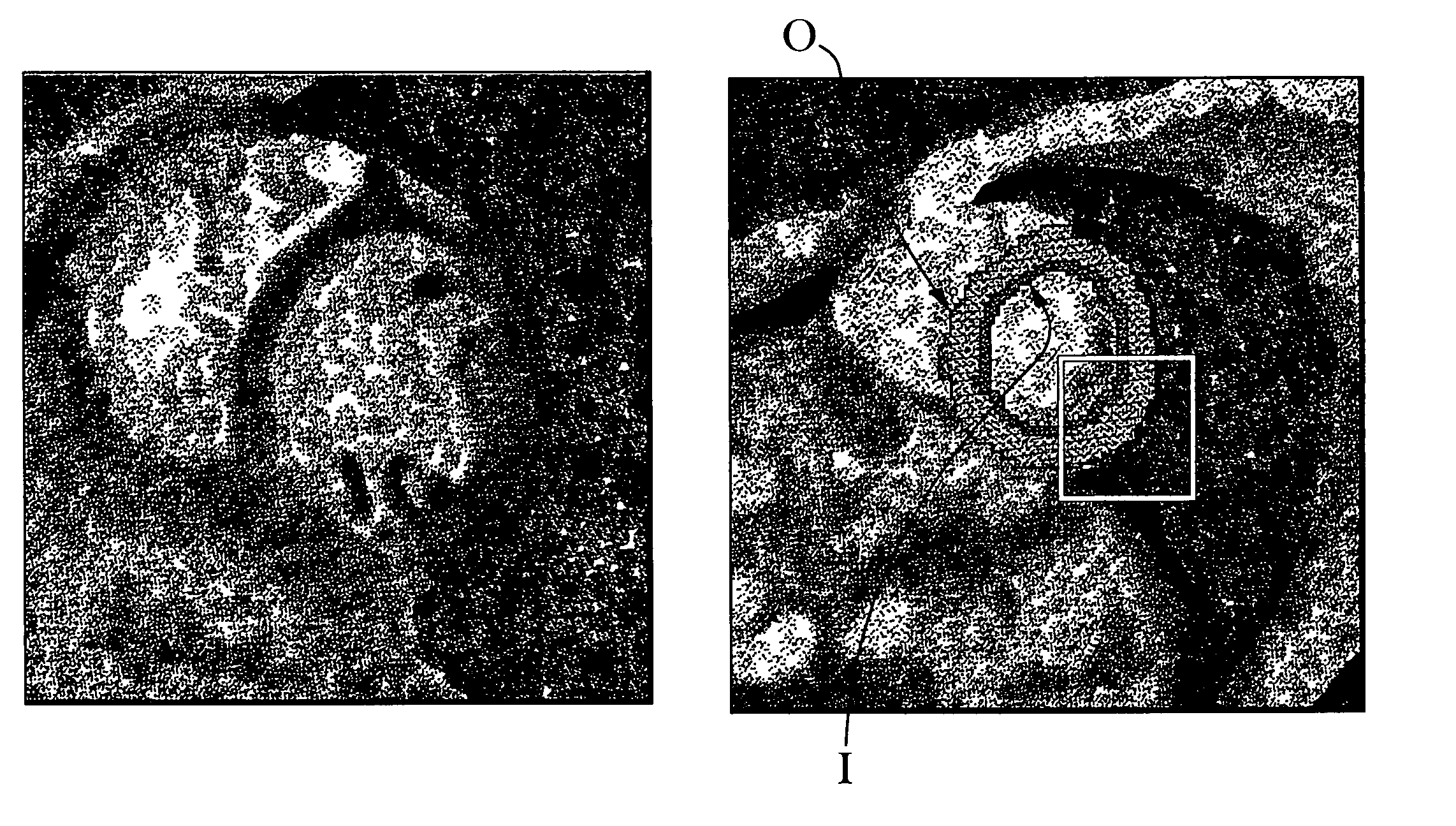
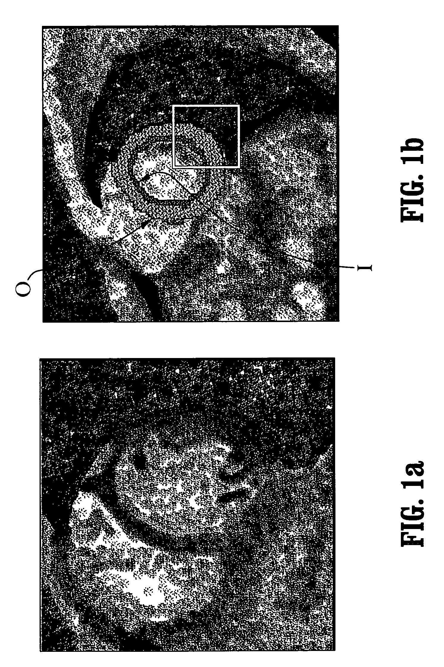
[0030]The present invention is generally directed to cardiac segmentation for MR imagery. An objective is to recover the left ventricle and, in particular, the myocardium that is the area between the epicardium (i.e., the inner most layer of the pericardium (sac that surround heart) and the endocardium (the thin endothelial membrane lining the cavities of the heart.) The context of a segmentation application according to the invention is shown in FIG. 1.

[0031]FIG. 1a is an exemplary 2-d image of the heart, and in particular, a 2-d slice of the heart showing a cross-section of the left ventricle. The dark ring in FIG. 1 depicts the myocardium (muscle) that constitutes the wall of the left ventricle. In accordance with a preferred embodiment of the present invention, a segmentation method is based on two evolving interfaces as shown in FIG. 1b, the endocardium [∂RI—inner contour] or inner wall (denoted I) of the myocardium and the epicardium [∂RO—outer contour] or the outer wall (deno...


PUM


Abstract

A system and method for segmenting cardiac images and, in particular, segmenting the left ventricle of the heart using a contour propagation model that integrates visual information and anatomical constraints. The visual information comprises a gradient vector flow-based boundary component and a region component that separates the cardiac contours/regions according to their global intensity properties that reflect the different tissue properties. The anatomical constraints couple the propagation of cardiac contours according to their relative distance. The propagation model comprises a weighted integration of a boundary segmentation model, a region model and coupling function.

Description

CROSS-REFERENCE TO RELATED APPLICATION[0001]This application claims priority to U.S. Provisional Patent Application Ser. No. 60 / 292,445, filed on May 17, 2001.TECHNICAL FIELD[0002]The present invention is generally directed to a system and method for segmenting cardiac images. In particular, the invention is directed to a system and method for segmenting the left ventricle of the heart using a contour propagation model that integrates visual information and anatomical constraints. The visual information comprises a gradient vector flow-based boundary component, and a region component that separates the cardiac contours / regions according to their global intensity properties that reflect the different tissue properties. The anatomical constraints couple the propagation of cardiac contours according to their relative distance.BACKGROUND[0003]Medical Image Processing is a growing application domain. Magnetic Resonance Imaging (MRI) may be used to provide a time-varying three dimensional...

Claims


Application Information

Patent Timeline
IPC IPC(8): G06K9/00G01R33/54A61B5/00A61B5/055G06T5/00G06T7/20
CPCG06T7/0083G06T7/0089G06T7/2006G06T2207/30048G06T2207/10088G06T2207/20161G06T7/12G06T7/149G06T7/215
Inventor PARAGIOS, NIKOLAOSRAMESH, VISVANATHAN
Owner SIEMENS CORP
Why Eureka
Try Eureka