
Forceps with spring loaded end effector assembly

a technology of end effectors and forceps, which is applied in the field of endoscopic surgical instruments, can solve the problems of not being able to safely coagulate arteries, surgeons can have difficulty suturing vessels or performing other traditional methods of controlling bleeding, and abandoning the benefits of laparoscopy, etc., and achieves the effect of sealing tissue upon application of electrosurgical energy

Active Publication Date: 2007-03-27
COVIDIEN AG
View PDF454 Cites 595 Cited by
  • Summary
  • Abstract
  • Description
  • Claims
  • Application Information

AI Technical Summary

Benefits of technology

This solution provides consistent and reliable sealing and separation of tissue along the sealing line, reducing the risk of mechanical failure and improving precision, allowing for effective sealing of larger vessels within the constraints of endoscopic procedures.

Problems solved by technology

Smaller cannulas are usually preferred, which, as can be appreciated, ultimately presents a design challenge to instrument manufacturers who must find ways to make surgical instruments that fit through the particularly-sized cannulas.
However, due to space limitations surgeons can have difficulty suturing vessels or performing other traditional methods of controlling bleeding, e.g., clamping and / or tying-off transected blood vessels.
However, if a larger vessel is severed, it may be necessary for the surgeon to convert the endoscopic procedure into an open-surgical procedure and thereby abandon the benefits of laparoscopy.
The article states that it is not possible to safely coagulate arteries with a diameter larger than 2 to 2.5 mm.
Below this range, the seal may shred or tear and above this range the lumens may not be properly or effectively sealed.
Various known electrosurgical instruments and methods may occasionally be able to seal larger vessels using an appropriate electrosurgical power curve, coupled with an instrument capable of applying a large closure force to the vessel walls, however, these instruments and methods rarely provide consistent and accurate vessel sealing.
Thus, coagulation of small vessels is sufficient to permanently close the vessels but not necessarily seal the vessels.
Many known electrosurgical instruments include blade members or shearing members which simply cut tissue in a mechanical and / or electromechanical manner and are relatively ineffective for vessel sealing purposes.
Other instruments rely on clamping pressure alone to procure proper sealing thickness and are not designed to take into account gap tolerances and / or parallelism and flatness requirements which are parameters which, if properly controlled, can assure a consistent and effective tissue seal.
For example, it is known that it is difficult to adequately control thickness of the resulting sealed tissue by controlling clamping pressure alone for either of two reasons: 1) if too much force is applied, there is a possibility that the two poles will touch and energy will not be transferred through the tissue resulting in an ineffective seal; or 2) if too low a force is applied the tissue may pre-maturely move prior to activation and sealing and / or a thicker, less reliable seal may be created.
This presents a challenge because the jaw members are typically affixed with pins which are positioned to have a small moment arms with respect to the pivot of each jaw member.
A large force, coupled with a small moment arm, is undesirable because the large forces may shear the pins.
Increasing the closure forces between electrodes may have other undesirable effects, e.g., it may cause the opposing electrodes to come into close contact with one another which may result in a short circuit and a small closure force may cause pre-mature movement of the issue during compression and prior to activation.
As can be appreciated, this additional step may be both time consuming (particularly when sealing a significant number of vessels or tissue) and may contribute to imprecise separation of the vessels or tissue along the sealing line due to the misalignment or misplacement of the severing instrument along the center of the sealing line.
It is believed that these wire-like jaw frames are not designed to seal tissues since, by and large, the jaw frames do not provide the forces necessary to effect consistent and accurate vessel sealing.

Method used


Image

  • Forceps with spring loaded end effector assembly
  • Forceps with spring loaded end effector assembly
  • Forceps with spring loaded end effector assembly

Examples


Embodiment Construction

[0036]Detailed embodiments of the presently disclosed instruments, devices and systems will now be described in detail with reference to the drawing figures wherein like reference numerals identify similar or identical elements. In the drawings and in the description which follows, the term “proximal”, as is traditional, will refer to the end of the instrument, device and / or system which is closest to the operator while the term “distal” will refer to the end of the instrument, device and / or system which is furthest from the operator.

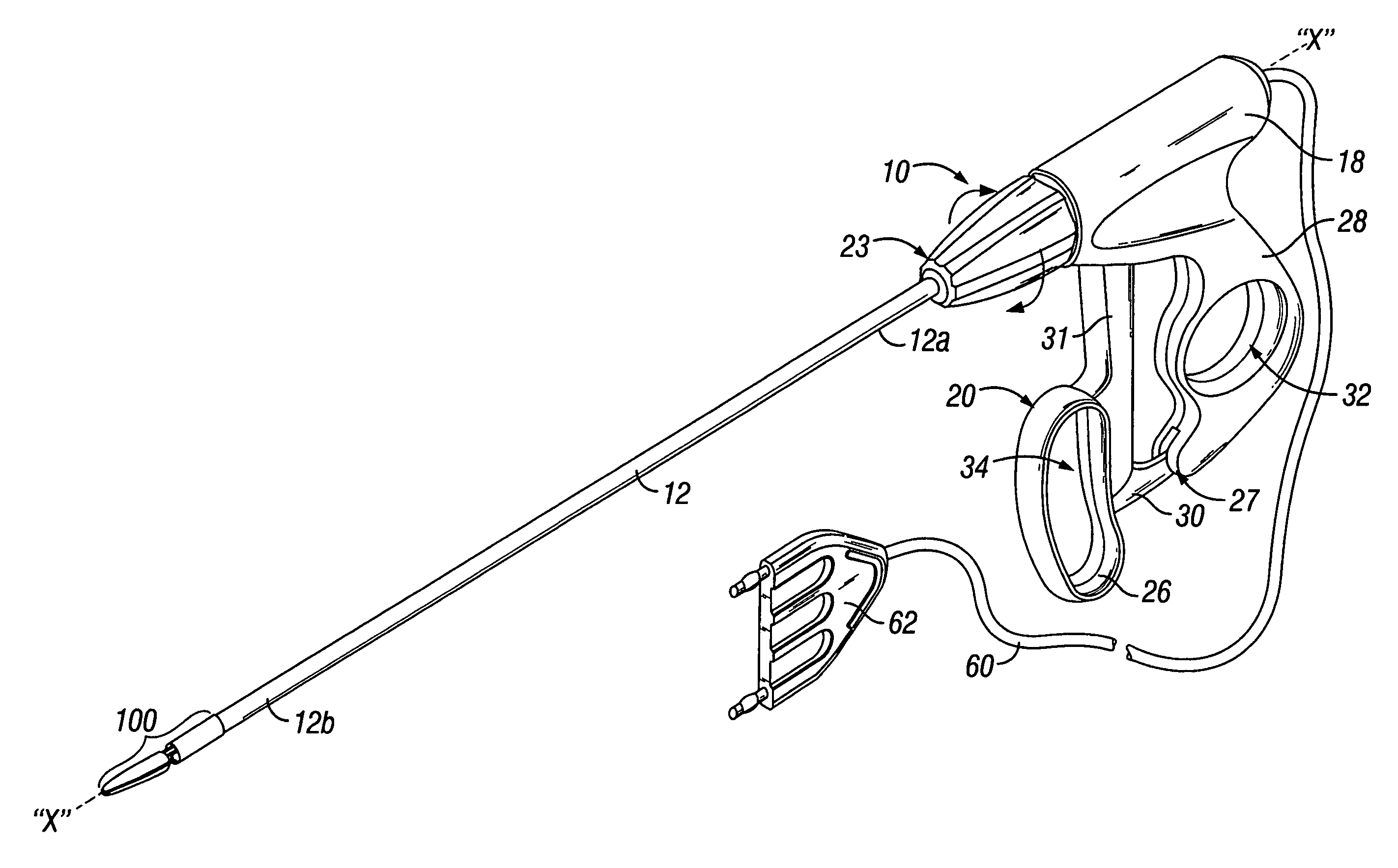
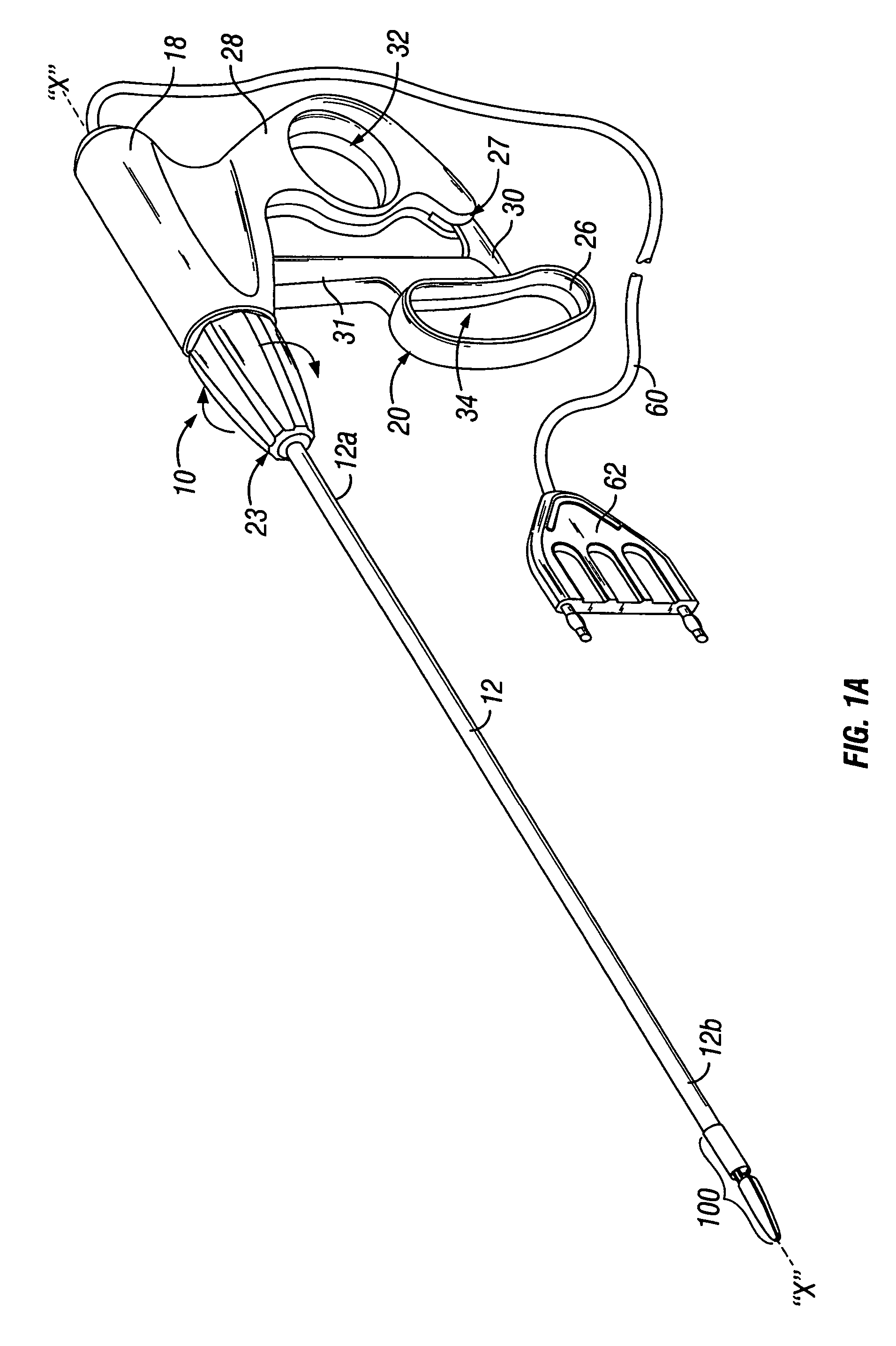
[0037]Referring to FIG. 1A, an endoscopic instrument according to one embodiment of the present disclosure is designated generally as reference numeral 10. Endoscopic instrument 10 includes an elongated shaft 12 which is mechanically coupled to a handle assembly 18 at a proximal end 12a thereof and a distal end 12b which is mechanically coupled to an end effector assembly 100. Elongated shaft 12 is preferably hollow and includes internal electrical conn...


PUM


Abstract

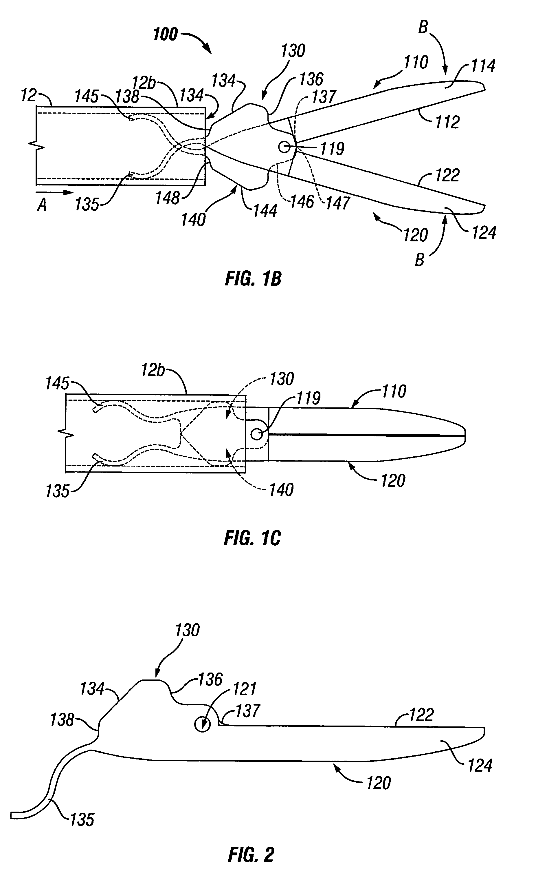
An endoscopic forceps for treating tissue includes a handle assembly having a elongated shaft extending therefrom which is movable relative to the handle assembly by an actuator. The forceps also including an end effector assembly attached to a distal end of the shaft which has first and second jaw members which are movable about a pivot from a first position in spaced relation relative to one another to a second position for clamping tissue. Each of the jaw members include: an insulative outer housing having an electrically conductive, inwardly facing tissue contacting surface and a proximal flange with a camming surface. The proximal flange being provided with a spring-like tail which mounts within the distal end of the elongated shaft to bias the jaw members in an open configuration. The actuator is movable relative to the handle to actuate the elongated shaft to cam the proximal flanges to pivot the jaw members toward the second position to clamp tissue.

Description

BACKGROUND[0001]1. Technical Field[0002]The present disclosure relates to endoscopic surgical instruments and, more particularly, to an end effector assembly for use in connection with endoscopic instruments for grasping, sealing, dividing and / or dissecting tissue.[0003]2. Background of Related Art[0004]A hemostat or forceps is a simple pliers-like tool which uses mechanical action between its jaws to constrict vessels and is commonly used in open surgical procedures to grasp, dissect and / or clamp tissue. Electrosurgical forceps utilize both mechanical clamping action and electrical energy to effect hemostasis by heating the tissue and blood vessels to coagulate, cauterize and / or seal tissue.[0005]Over the last several decades, more and more surgeons are complimenting traditional open methods of gaining access to vital organs and body cavities with endoscopes and endoscopic instruments which access organs through small puncture-like incisions. Endoscopic instruments are inserted int...

Claims


Application Information

Patent Timeline
Patent Type & Authority Patents(United States)
IPC IPC(8): A61B18/04
CPCA61B17/29A61B18/1442A61B2017/2929A61B2017/2944A61B2017/2937A61B2017/2939A61B2017/2933A61B2017/2845A61B2017/2926
Inventor DUMBAULD, PATRICK L.
Owner COVIDIEN AG
Learn More