Joint analysis using ultrasound

a joint analysis and ultrasound technology, applied in the field of quantitative joint measurements using ultrasound, can solve the problems of not being accepted as joint measurement tools, ultrasound has not lived up to its potential, and the observation of joint structures with ultrasound

Inactive Publication Date: 2007-04-17
JOINTVIEW
View PDF37 Cites 62 Cited by
  • Summary
  • Abstract
  • Description
  • Claims
  • Application Information

AI Technical Summary

Problems solved by technology

Observation of joint structures with ultrasound, though described in many articles in the past decades, has failed to become an accepted joint measurement tool.
Yet, for all its advantages, ultrasound has not lived up to its potential.
Ultrasound has apparently not gained widespread use for assessing joints beyond imaging, in spite of its apparent suitability.

Method used


Image

  • Joint analysis using ultrasound
  • Joint analysis using ultrasound
  • Joint analysis using ultrasound

Examples


Embodiment Construction

Overview of Osteoarthritis

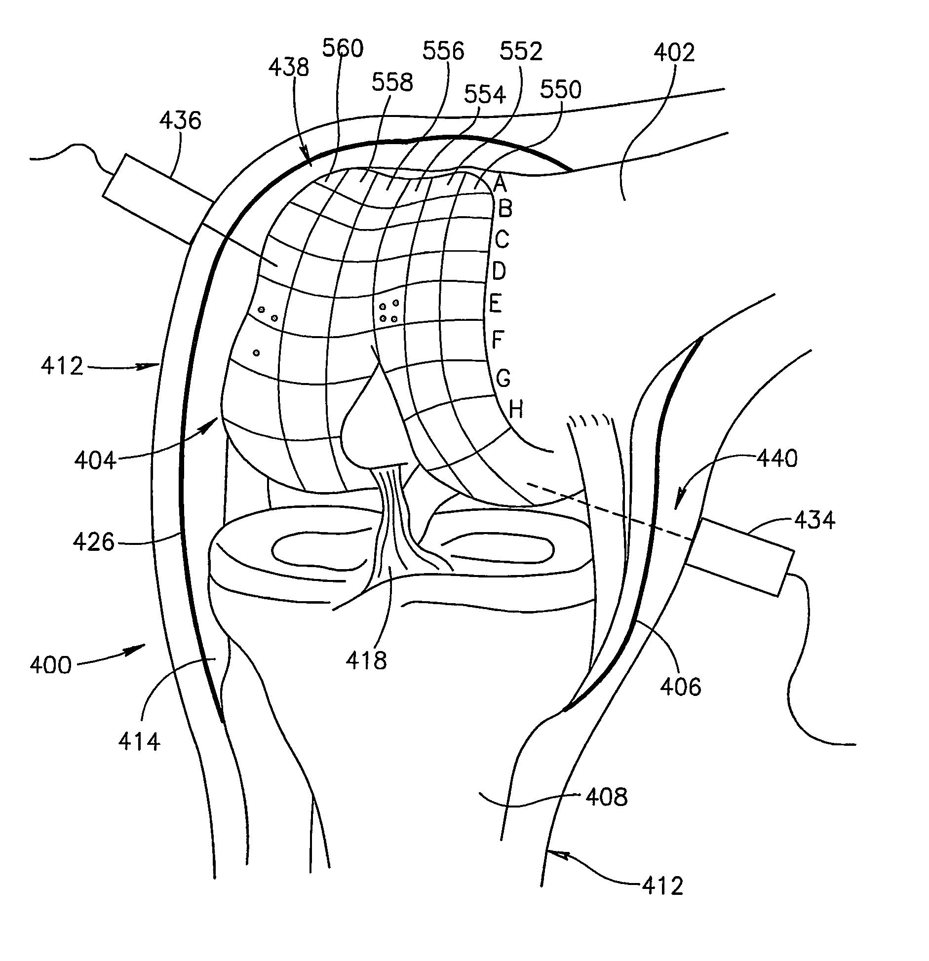
[0091]FIG. 1 illustrates anatomy for the purpose of making quantitative measurements of joint, for example, to indicate or aid in diagnosis of and / or track osteoarthritis (OA). OA is a major cause of pain in joints such as joint 100. Bones, such as a tibia 108 and a femur 102, in a healthy joint, fit closely together. End of femur 102 is coated with an area of cartilage 104 and end of tibia 108 is coated with an area of cartilage 116 that provide a cushion during motion.

[0092]In the early stages of OA, in addition to inflammation and swelling of joint capsule 106, a plurality of focal blisters 144 may form, such as in cartilage surface 104 of femur 106. As OA progresses, thinning occurs in a cartilage surface, such as cartilage surface 116 of tibia 108. An area of eburnation 140 can occur in cartilage surface 116, where cartilage 116 is completely worn away to expose bone 108. Cartilage 116 does not have its own blood supply so damaged cartilage 116 does no...


PUM


Abstract

A method is provided for generating at least one quantitative measure of a joint comprising sending an acoustic signal to at least a part of a joint, receiving said acoustic signal after modification by said joint, analyzing the received acoustic signal with a computer, and generating, from said computer, one or more of a quantitative surface, quantitative volumetric and quantitative measurements of the physical properties of the joint or a portion thereof.

Description

RELATED APPLICATIONS[0001]The present application is a US National Phase of PCT. Application No. PCT / IL01 / 00763, filed on Aug. 16, 2001.FIELD OF THE INVENTION[0002]The present invention relates to quantifying joint measurements using ultrasound.BACKGROUND OF INVENTION[0003]Observation of joint structures with ultrasound, though described in many articles in the past decades, has failed to become an accepted joint measurement tool. As early as 1983, ultrasound was suggested as a means of observing joint cartilage. As an example, Alex M. Aisen, MD et. al. Radiology Vol. 153 Number 3, December 1983, “Sonographic Evaluation of the Cartilage of the Knee,” demonstrates the use of ultrasound in observing “changes in the surface characteristics and thickness of cartilage”. This ultrasound method relies on subjectively evaluating an ultrasound image.[0004]Ultrasound has the potential to be useful in observing some joint structures besides cartilage. According to Iagnocco, et al.Scand. J Rheu...

Claims


Application Information

Patent Timeline
Patent Type & Authority Patents(United States)
IPC IPC(8): A61B8/00A61B5/107A61B8/08A61F2/00A61F2/30
CPCA61B5/107A61B5/4514A61B8/0858A61B8/0875A61B5/4528A61B5/6847A61B8/587A61F2/30756A61F2002/30705A61F2002/307
Inventor TSOREF, LIAT
Owner JOINTVIEW
Why Eureka
Try Eureka