Viewing system having means for processing a sequence of ultrasound images for performing a quantitative estimation of flow in a body organ

a viewing system and ultrasound technology, applied in the field of viewing systems, can solve the problems of reverberating against the heart walls, underestimating the extent of the jet, and eccentricity of the regurgitant j

Inactive Publication Date: 2007-06-12
KONINKLIJKE PHILIPS ELECTRONICS NV
View PDF0 Cites 88 Cited by
  • Summary
  • Abstract
  • Description
  • Claims
  • Application Information

AI Technical Summary

Benefits of technology

[0007]The invention has for an object to provide a viewing system associated to an ultrasound examination apparatus and an image processing method for performing an automatic quantitative estimation of a flow through a body organ, from a sequence of three-dimensional (3-D) ultrasound color flow images. According to the invention, a 3-D ultrasound technique is used for acquiring a sequence of 3-D color flow images of a flow through an orifice, for instance using a 3-D color Doppler technique, within an interval of time. Then, a map of isovelocity surfaces is constructed by velocity segmentation related to the blood flow at the vicinity of said orifice, issuing through the orifice, which permits of estimating both the blood velocity at a given surface and the actual volume delimited by said isovelocity surface. The peak velocity of the regurgitant jet of blood through the orifice, in the opposite direction, is also measured using a pulse wave Doppler technique. These measured data permit of calculating the surface of the orifice. In particular, for estimating the severity of the mitral regurgitation through the heart valve, the Surface Of Regurgitation (SOR) can be obtained by calculations based on the real volume corresponding to the real PISA.

Problems solved by technology

However, these measurements, even though they are based on the maximal demonstrable regurgitant jet, tend to underestimate the extent of the jet.
Moreover, the regurgitant jet can be eccentric or asymmetric, and can reverberate against the heart walls, which is the case in most severe mitral regurgitations for instance, and which does not permit of assessing the actual mitral regurgitation correctly.
Hence, on the one hand, the known 2-D measurements do not permit of quantifying said mitral regurgitation accurately.
However, on the other hand, the publication by HIROKO FUJI et alii, in reference, which is based on geometrical assumption limited to a very strict hemielliptic shape of the isovelocity surface of the 3D color flow of the blood issuing the mitral valve, does not permit of reaching sufficiently accurate results.

Method used


Image

  • Viewing system having means for processing a sequence of ultrasound images for performing a quantitative estimation of flow in a body organ
  • Viewing system having means for processing a sequence of ultrasound images for performing a quantitative estimation of flow in a body organ
  • Viewing system having means for processing a sequence of ultrasound images for performing a quantitative estimation of flow in a body organ

Examples


Embodiment Construction

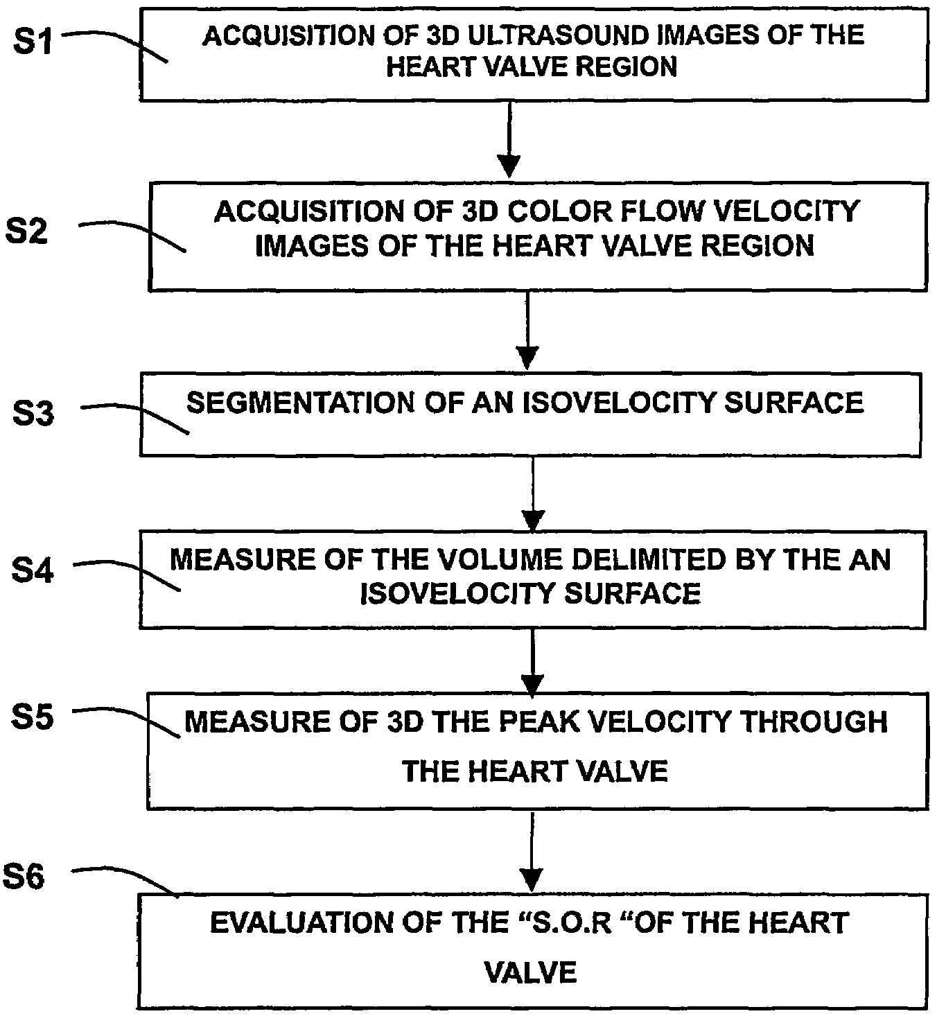
[0014]The invention relates to a viewing system for performing a quantitative estimation of a flow in a body organ. In particular, the invention relates to a medical viewing system and to an image processing method for performing an automatic quantitative estimation of the blood flow through the heart valves, and / or of the regurgitant jet, from a sequence of 3-D color flow images.

[0015]The method can be carried out using reconstructed or real-time 3D echocardiography, the images being formed using a trans-thoracic or a trans-esophageal probe. The method of the invention can also be applied to a sequence of 3-D images of other organs of the body that can be formed by ultrasound systems or ultrasound apparatus, or by other medical imaging systems known of those skilled in the art.

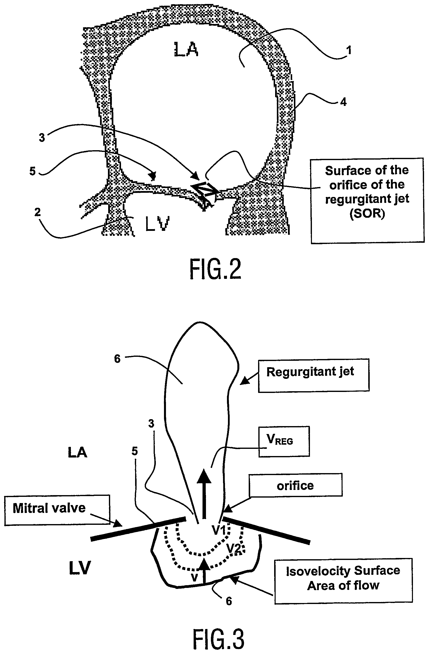
[0016]In the example described hereafter, severity of the cardiac regurgitant jet between the left heart atrium and the left heart ventricle is assessed from a sequence of 3-D Doppler color flow images.

[0017]...


PUM


Abstract

A medical ultrasound viewing system for processing a sequence of three-dimensional (3-D) ultrasound images far performing a quantitative estimation of a flow through a body organ comprising means for performing steps of acquiring a sequence of 3D color flow images, of said flow; assessing the flow velocity values in the 3D images, constructing isovelocity surfaces (6) by segmentation of the flow velocity values; computing the volume (Vol) delimited by the isovelocity surfaces; and using the flow velocity value (V) and the volume (Vol) computed from a segmented surface for computing the surface of an orifice (3) of the organ through which the flow propagates. The viewing system further comprises means for performing steps of measuring the peak velocity (VREG) of said flow through said orifice; computing the surface of the orifice (SOR) through which the flow propagates as a function of the flow velocity value (V) at an isovelocity surface upstream the flow propagation with respect to said orifice, the volume (Vol) computed from said segmented isovelocity surface, and the peak velocity of the flow through said orifice. The surface is given by the formula: SOR=Vol. V / VREG. The system can be applied to the assessment of the surface of regurgitation of the mitral jet.

Description

CROSS-REFERENCE TO RELATED APPLICATIONS[0001]This application claims priority from the pending International Patent Application PCT / IB02 / 05356 filed on Dec. 11, 2002, and from the pending European Patent Application 01403392.2 filed on Dec. 28, 2001.FIELD OF THE INVENTION[0002]The invention relates to a viewing system having means for processing a sequence of three-dimensional (3-D) ultrasound images for performing a quantitative estimation of a flow in a body organ. In particular, the invention relates to a medical viewing system and to an image processing method for performing an automatic quantitative estimation of the blood flow through the heart valves, and / or of the regurgitant jet, from a sequence of 3-D color flow images.[0003]The invention particularly finds an application in the field of medical imaging in cardiology using ultrasound medical apparatus and / or viewing systems.BACKGROUND OF THE INVENTION[0004]A method for assessing mitral regurgitation is already known from t...

Claims


Application Information

Patent Timeline
Patent Type & Authority Patents(United States)
IPC IPC(8): A61B8/00A61B8/06A61B8/12G01S15/89
CPCA61B8/06G01S15/8979A61B8/0883G01S15/8993
Inventor JACOB, MARIEGERARD, OLIVIERCOLLET-BILLON, ANTOINE
Owner KONINKLIJKE PHILIPS ELECTRONICS NV
Why Eureka
Try Eureka