Automated method of patient recognition using chest radiographs
What is AI technical title?
AI technical title is built by Patsnap AI team. It summarizes the technical point description of the patent document.
a chest radiograph and automatic method technology, applied in image enhancement, instruments, healthcare informatics, etc., can solve the problems of difficult to find such filing errors, difficult to re-file images in the correct location of the pacs server, and inability to recognize patients
Inactive Publication Date: 2007-07-31
UNIVERSITY OF CHICAGO
View PDF9 Cites 6 Cited by
Summary
Abstract
Description
Claims
Application Information
AI Technical Summary
This helps you quickly interpret patents by identifying the three key elements:
Problems solved by technology
Method used
Benefits of technology
Benefits of technology
[0015]Accordingly, an object of this invention is to provide a method and system that discovers and prevents filing errors in archiving and retrieving images in the PACS environment.
Problems solved by technology
If a patient's information associated with an acquired image does not match the correct information on the patient, a filing error will occur in the PACS environment.
The main reasons for filing errors are related to human errors, such as incorrect input of patient information, accidental acquisition of radiographs of a wrong patient for a given examination, or occasionally imperfect design of the PACS [1][2].
Thus, the image may be assigned to a different patient name and may not be stored in the proper patient's folder.
It is generally difficult to find such filing errors.
Even if radiology personnel discover “wrong” images in the PACS server at a later date, it is difficult to re-file the image in the correct location in the PACS server.
Filing errors may create serious problems, e.g., retrieval failure for a specific image from the PACS server [1], or radiologists may interpret incorrect images for a given patient.
Although such serious errors do not occur frequently in clinical situations, it is known that filing errors mainly caused by human mistakes occur in the PACS environment [1][2].
Method used
Image
Click on the blue label to locate the original text in one second.
Reading with bidirectional positioning of images and text.
Examples
Embodiment Construction
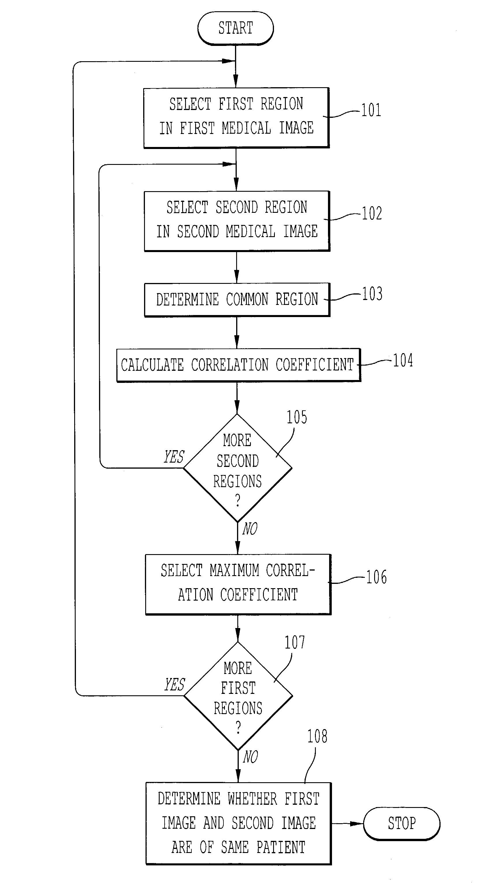
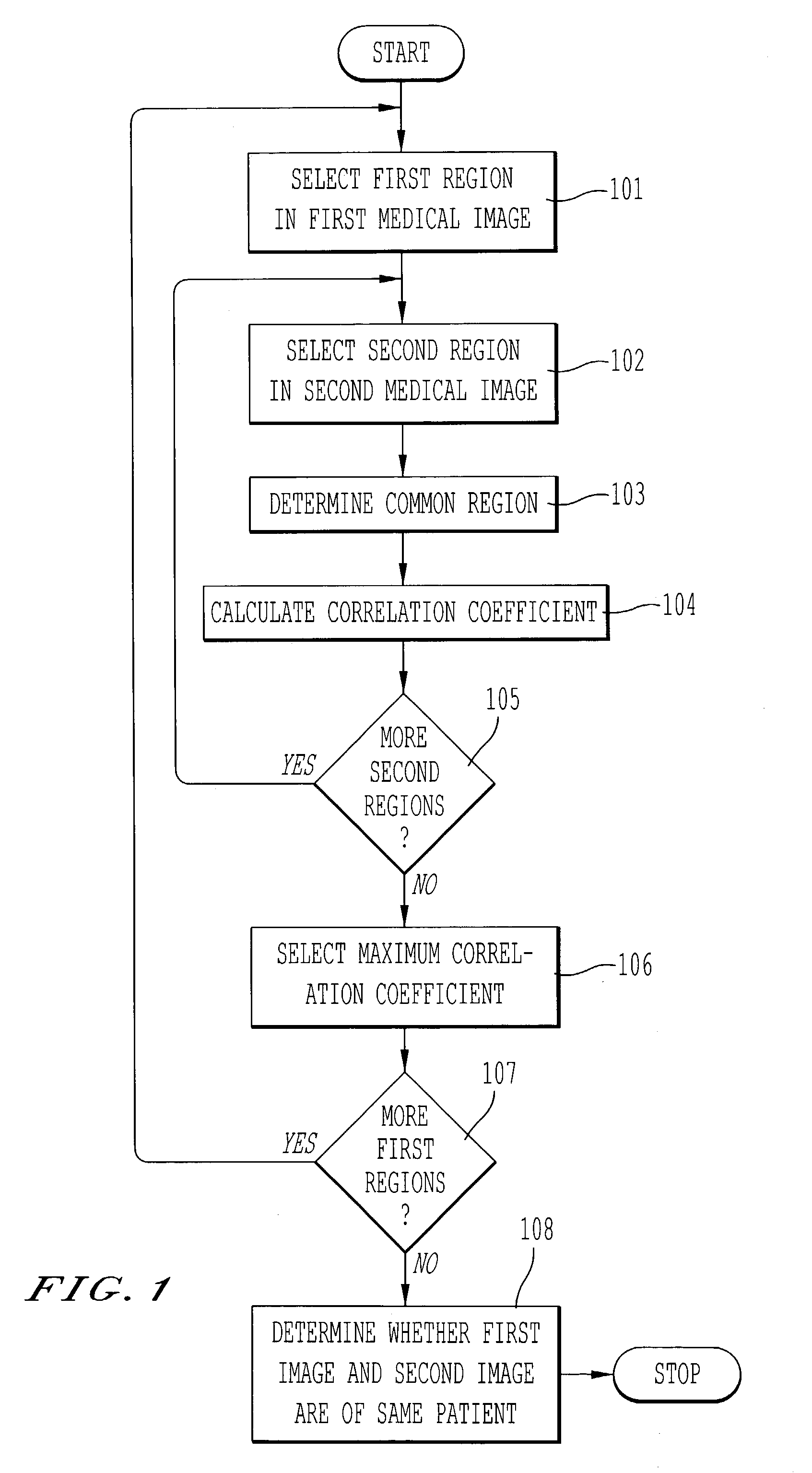
[0034]Referring now to the drawings, wherein like reference numerals designate identical or corresponding parts throughout the several views, FIG. 1 is a flowchart of a method for determining whether a first medical image and a second medical image are medical images of the same patient. In step 101, a first region is selected in the first medical image. The first region corresponds, for example, to one of a thoracic field, a cardiac shadow, a lung apex, a superior mediastinum, and a right lower lung in the first medical image. In step 102, a second region is selected within a search region of the second medical image. The search region is based on the first region selected in the first medical image. In step 103, a region common to the first region and the second region is determined based on a boundary of the first region and a boundary of the second region. Next, in step 104, a correlation coefficient is calculated based on image data from the first medical image in the common re...
PUM
Abstract
A method for determining whether a first medical image and a second medical image are medical images of the same patient, comprising selecting a first region in the first medical image; selecting a second region in the second medical image; determining a common region based on a boundary of the first region and a boundary of the second region; calculating a correlation coefficient based on image data from the first medical image in the common region and image data from the second medical image in the common region; and determining whether the first medical image and the second medical image are medical images of the same patient based on the correlation coefficient. Biological fingerprints from parts of chest radiographs such as thoracic fields, cardiac shadows, lung apices, superior mediastinum, and the right lower lung that includes the costophrenic angle, are used for the purpose of patient recognition and identification.
Description
CROSS-REFERENCE TO RELATED APPLICATIONS[0001]The present application is related to and claims priority to U.S. Provisional Application Ser. No. 60 / 428,939, filed Nov. 26, 2002. The contents of that application are incorporated herein by reference.STATEMENT REGARDING FEDERALLY SPONSORED RESEARCH[0002]The present invention was made in part with U.S. Government support under NIH Grant No. CA62625. The U.S. Government may have certain rights to this invention.BACKGROUND OF THE INVENTIONField of the Invention[0003]The present invention relates generally to systems and methods for computer-aided patient recognition and identification using biological fingerprints obtained from radiographs.[0004]The present invention also generally relates to computerized techniques for automated analysis of digital images, for example, as disclosed in one or more of U.S. Pat. Nos. 4,839,807; 4,841,555; 4,851,984; 4,875,165; 4,918,534; 5,072,384; 5,150,292; 5,224,177; 5,289,374; 5,319,549; 5,343,390; 5,359...
Claims
Application Information
Patent Timeline
Application Date:The date an application was filed.
Publication Date:The date a patent or application was officially published.
First Publication Date:The earliest publication date of a patent with the same application number.
Issue Date:Publication date of the patent grant document.
PCT Entry Date:The Entry date of PCT National Phase.
Estimated Expiry Date:The statutory expiry date of a patent right according to the Patent Law, and it is the longest term of protection that the patent right can achieve without the termination of the patent right due to other reasons(Term extension factor has been taken into account ).
Invalid Date:Actual expiry date is based on effective date or publication date of legal transaction data of invalid patent.

