
Anthropomorphic phantoms and method

a technology of phantoms and phantoms, applied in the field of ultrasonic imaging devices, can solve the problems of limited use of cadavers, animals, consenting patients, and not widely available training for cadavers, and achieve the effect of reducing the number of uses and not having the “look and feel” of human tissue during ultrasonic imaging procedures

Active Publication Date: 2007-08-14
CAE HEALTHCARE CANADA
View PDF22 Cites 91 Cited by
  • Summary
  • Abstract
  • Description
  • Claims
  • Application Information

AI Technical Summary

Benefits of technology

The phantom provides a realistic training tool that mimics human tissue, allowing for repeated use, stable at room temperature, and effectively simulates normal and abnormal anatomical structures, enhancing the training of healthcare workers in ultrasound imaging.

Problems solved by technology

Unfortunately, cadavers, animals, and consenting patients are not widely available for training.
One drawback with ultrasonic phantoms found in the prior art is that they do not have the “look and feel” of human tissue during an ultrasonic imaging procedure.
A second drawback is that they are not self-sealing after puncturing with a cannula or needle, thereby limiting their number of uses.
A third drawback is that they do not include internal structures designed to simulate normal and abnormal anatomical structures typically found during an ultrasonic imaging procedure on human tissue.
A fourth drawback is that they are susceptible to mold growth and quickly dry out when exposed to air for extended periods.

Method used


Image

  • Anthropomorphic phantoms and method
  • Anthropomorphic phantoms and method
  • Anthropomorphic phantoms and method

Examples


Embodiment Construction

)

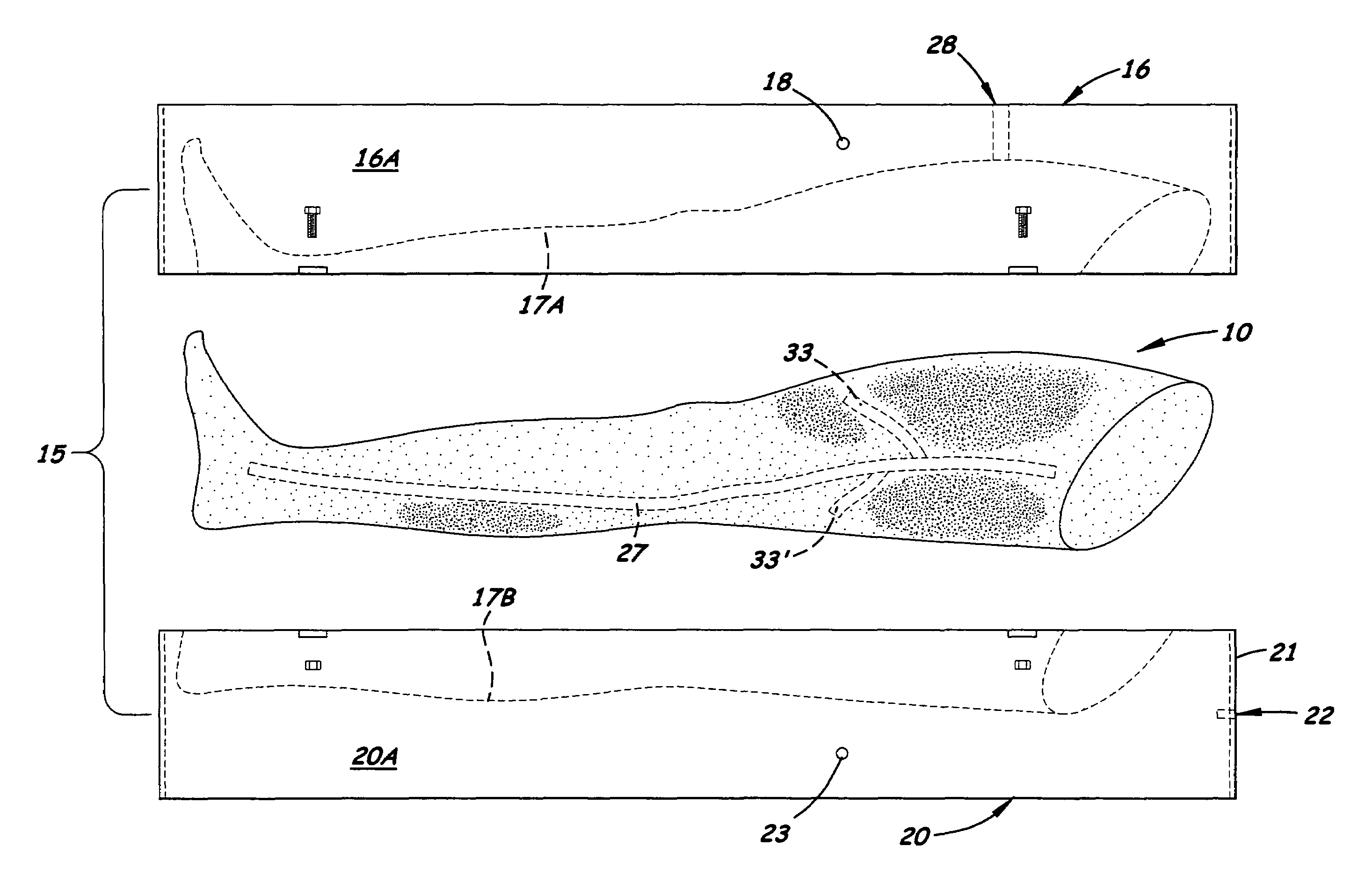
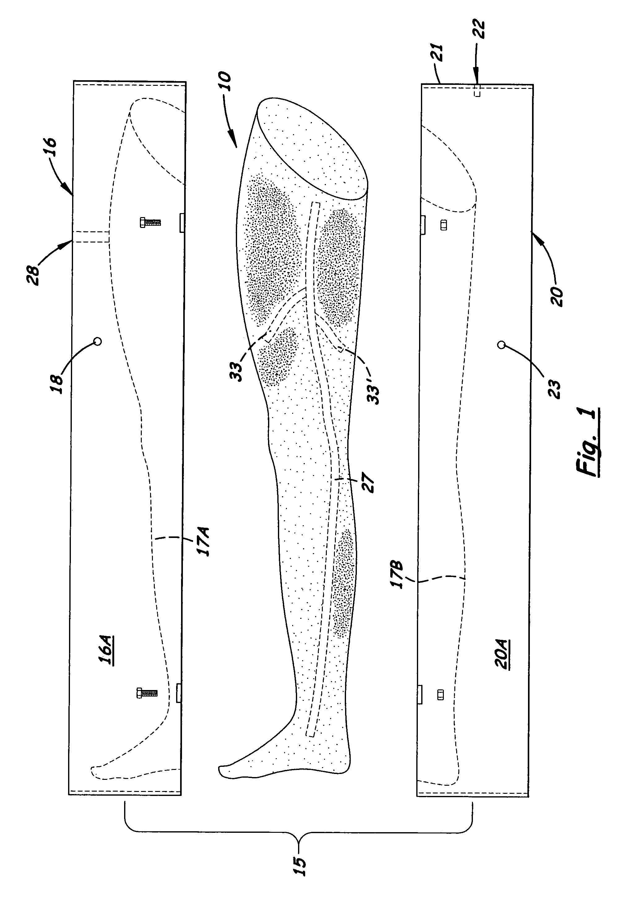
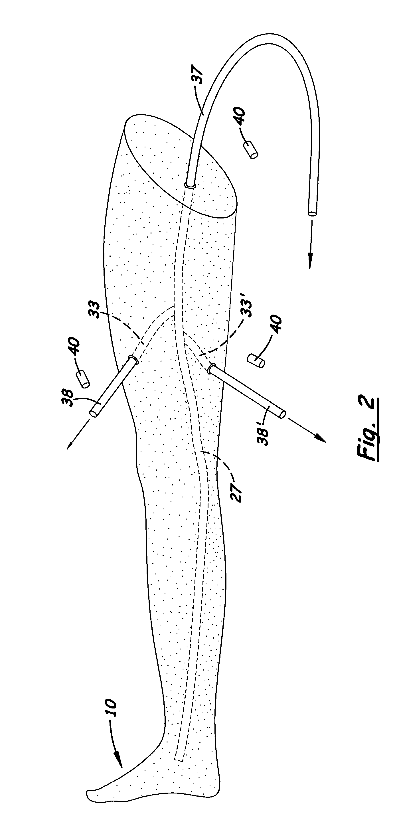
[0027]Referring to the FIGS., there is shown an anthropomorphic phantom 10 disclosed herein made of a chemical composition 11 that has the ‘look and feel’ of human tissue during an ultrasonic imaging procedure. The chemical composition 11 is made of moldable material. thereby enabling the phantom to be formed in a wide variety of different anatomical structures. Although the chemical composition 11 is substantially non-reflective during an ultrasonic image procedure, varying amounts of a scattering agent 25 may be added to the chemical composition 11 thereby enabling the manufacturer to adjust the sonographic characteristics of the chemical composition 11 to more closely mimic human tissue.

[0028]In the preferred embodiment, the chemical composition 11 are thermoplastic elastomers 12, 13 that are melted and then poured into a rigid primary mold 15. The thermoplastic elastomers 12, 13 are commercially available compositions comprised in part of highly plasticized styrene, ethylene, b...


PUM


Abstract

An anthropomorphic phantom used as a training tool by healthcare workers for learning ultrasonic imaging procedures. The phantom is made of a moldable, elastomeric tissue-simulating chemical composition that may be easily processed to create an anthropomorphic phantom: that has the “look and feel” and self-sealing characteristics of human tissue. During the mixing processes, scattering agents and pigments may be added to provide a phantom that simulates the sonographic characteristics on living tissue. The phantom body may contain objects and empty or liquid filled cavities and conduits that simulate normal internal structures and abnormal artifacts and conditions. In one embodiment, hollow rods are disposed longitudinally inside the primary mold that forms blood vessels inside the phantom. When the phantom is removed from the primary mold, the hollow rods are removed thereby forming a hollow conduit. Two plugs may be inserted into the ends of the conduit to hold a blood simulating liquid placed inside the conduit.

Description

BACKGROUND OF THE INVENTION[0001]1. Field of the Invention[0002]The present invention relates to the field of ultrasound imaging devices including diagnostic medical ultrasound, continuous wave Doppler, pulsed Doppler, duplex Doppler / imaging systems, color power Doppler, color-flow Doppler and methods for using them in medical imaging and other such applications. More particularly, this invention relates to phantoms used to assess the performance of ultrasound imaging devices, to measure quality assurance, and to teach ultrasound imaging and medical ultrasound guided techniques.[0003]2. Description of the Related Art[0004]Medical ultrasound imaging is used in a variety of different medical procedures. Among its applications is its use as a diagnostic testing modality and a method of guiding invasive procedures. Heretofore, ultrasound-imaging procedures have traditionally been performed by technicians and interpreted by physicians within radiology departments. Today, such procedures ...

Claims


Application Information

Patent Timeline
Patent Type & Authority Patents(United States)
IPC IPC(8): G09B23/28
CPCG09B23/286G09B23/303
Inventor KEEGAN, BRIAN
Owner CAE HEALTHCARE CANADA
Learn More