
Image-guided medical intervention apparatus and method

a medical intervention and imageguided technology, applied in the direction of radiation control devices, instruments, therapy, etc., can solve the problems of reducing the effectiveness of radiation therapy, generating degree of error by patient movement (whether voluntary), and unsuitable conventional medical imaging systems and methods

Active Publication Date: 2007-09-04
UNIVERSITY OF CHICAGO
View PDF26 Cites 94 Cited by
  • Summary
  • Abstract
  • Description
  • Claims
  • Application Information

AI Technical Summary

Benefits of technology

The present invention provides an image-guided radiotherapy apparatus and method that allows for precise targeting of tumors and minimizes damage to healthy tissues surrounding them. The apparatus includes a gantry, a radiation source, and a gamma ray photon imaging device. The gamma ray photon imaging device can detect gamma ray photons emitted from within a patient's body and generate an image based on the detected photons. The position and orientation of the radiation source and the gamma ray photon imaging device can be adjusted to change the trajectory of the beam of radiation and obtain different images of the patient. This allows for real-time monitoring and adjustment of the treatment plan, improving the accuracy of the therapy and minimizing the risk of complications.

Problems solved by technology

In many cases, however, conventional medical imaging systems and methods are either unsuitable for many types of medical interventions or are limited in application in one or more significant manners.
After medical images of a patient have been taken in order to determine the location of one or more body areas requiring radiation therapy, some degree of error is generated by patient movement (whether voluntary or involuntary).
This discrepancy can reduce the effectiveness of the radiation therapy, and in some cases can require that such therapy be prolonged and / or reduced in dose.
As another example, many types of medical interventions cannot be performed in conjunction with conventional medical imaging systems without moving the patient or moving the medical intervention apparatus to gain access to the patient.
Such movement can require interruption of medical imaging, compromise the quality of the medical images, reduce the effectiveness of the medical intervention, or have other undesirable effects.
For example, many types of computed tomography (CT) imaging, magnetic resonance imaging (MRI), and Positron Emission Tomography (PET) imaging devices at least partially enclose a patient, or otherwise significantly limit free access to the patient for medical intervention procedures.
Such movement typically results in one or more of the undesirable results mentioned above.

Method used


Image

  • Image-guided medical intervention apparatus and method
  • Image-guided medical intervention apparatus and method
  • Image-guided medical intervention apparatus and method

Examples


Embodiment Construction

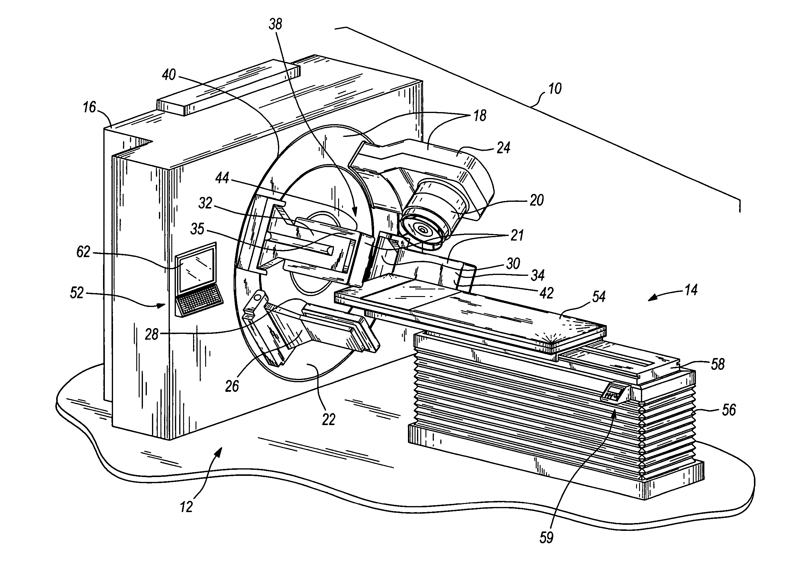
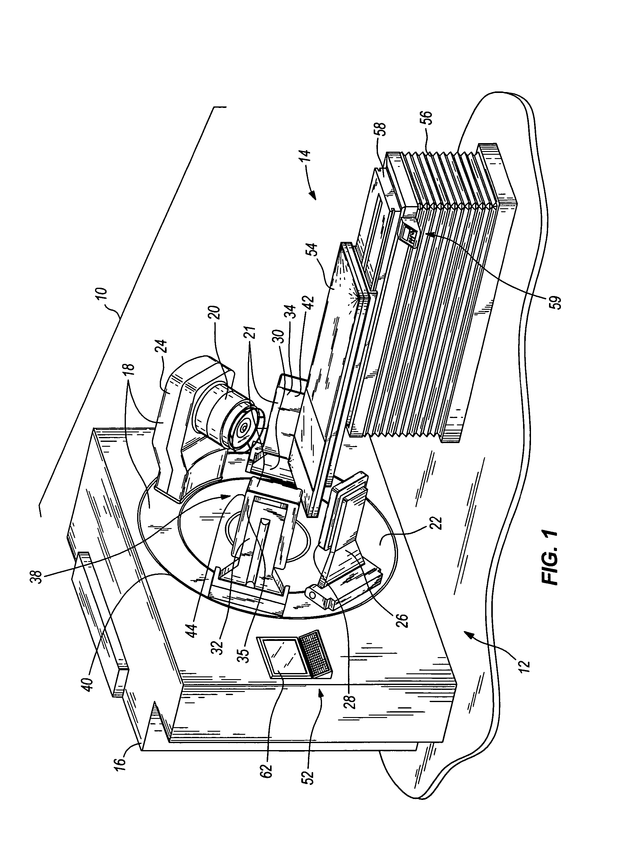
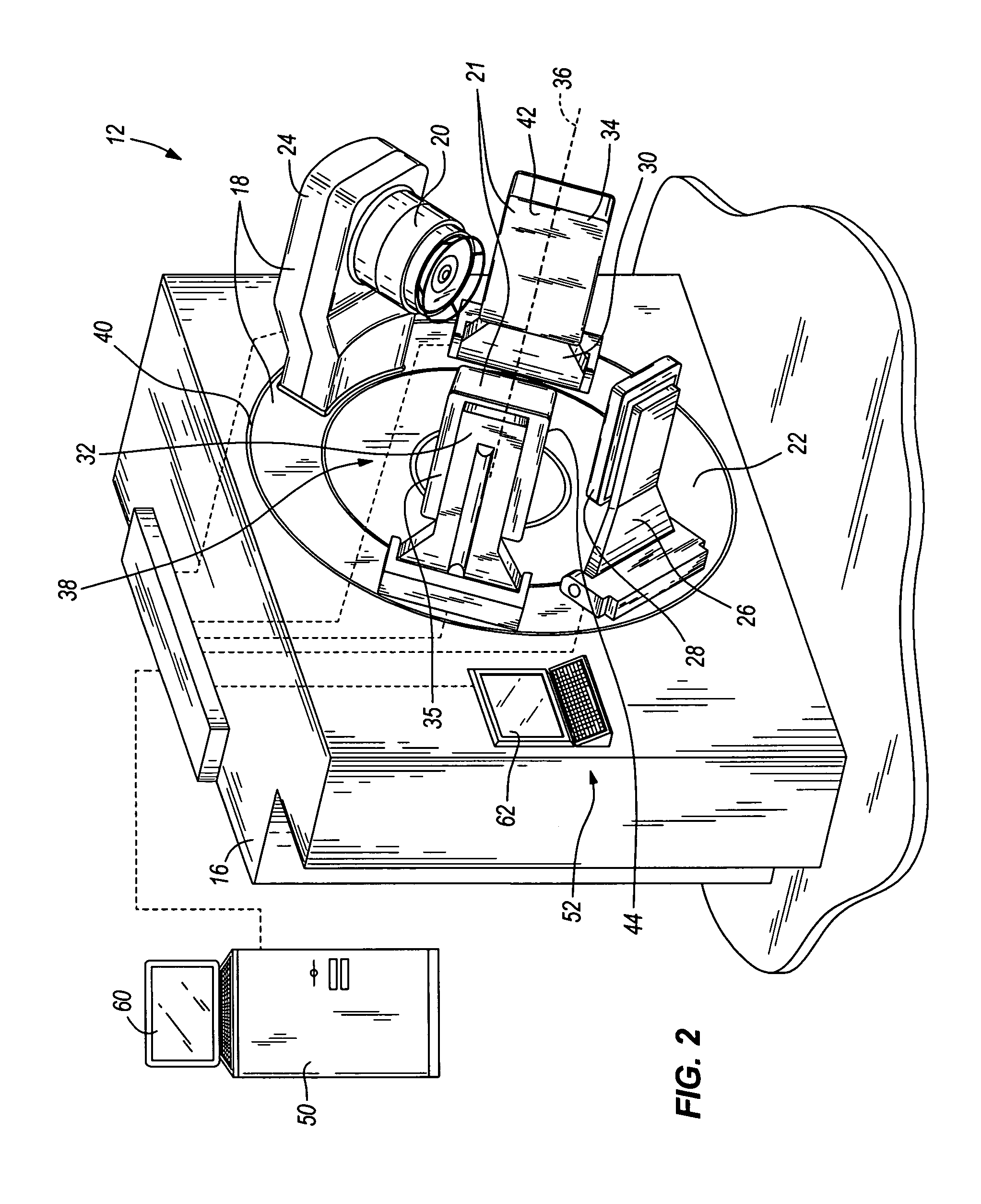
[0019]An image-guided radiotherapy apparatus according to an embodiment of the present invention is illustrated by way of example in FIGS. 1 and 2. The illustrated apparatus (indicated generally at 10) comprises a radiotherapy and imaging assembly 12 and a patient support 14. The patient support 14 is adapted to support a patient (i.e., a human or animal) in a position with respect to the radiotherapy and imaging assembly 12 during therapy and imaging procedures as described in greater detail below.

[0020]The radiotherapy and imaging assembly 12 illustrated in FIGS. 1 and 2 comprises a housing 16, a gantry 18 movable with respect to the housing 16, a radiotherapy accelerator 20 coupled to the gantry 18, and a PET imaging device 21 coupled to the gantry 18. As will be described in greater detail below, the gantry 18 is movable to different positions for purposes of administering radiotherapy and for PET image acquisition.

[0021]The gantry 18 in the illustrated embodiment of FIGS. 1 and...


PUM


Abstract

In some embodiments, an image-guided radiotherapy apparatus and method is provided in which a radiotherapy radiation source and a gamma ray photon imaging device are positioned with respect to a patient area so that a patient can be treated by a beam emitted from the radiotherapy apparatus and can have images taken by the gamma ray photon imaging device. Radiotherapy treatment and imaging can be performed substantially simultaneously and / or can be performed without moving the patient in some embodiments. The gamma ray photon imaging device can be coupled and movable with respect to any part of a building structure, can be located on a portable frame movable to and from the radiotherapy radiation source and patient, or can take other forms. In some embodiments, the gamma ray photon imaging device can be used for imaging in connection with other types of medical interventions.

Description

BACKGROUND OF THE INVENTION[0001]In virtually all fields of medicine, the pace of technological developments continues to drive the need for improved medical imaging devices and methods. Such devices and methods are commonly employed in conjunction with many different types of medical interventions, such as radiotherapy, surgery, biopsies, and the like.[0002]In many cases, however, conventional medical imaging systems and methods are either unsuitable for many types of medical interventions or are limited in application in one or more significant manners. By way of example only, the effectiveness of conventional radiotherapy systems and methods is typically a function of medical image accuracy. After medical images of a patient have been taken in order to determine the location of one or more body areas requiring radiation therapy, some degree of error is generated by patient movement (whether voluntary or involuntary). As a result, the precise locations of the body areas are often ...

Claims


Application Information

Patent Timeline
Patent Type & Authority Patents(United States)
IPC IPC(8): G01T1/24
CPCA61N5/1049G01T1/1641G01T1/166A61N2005/1052A61N2005/1061A61N5/1065
Inventor PELIZZARI, CHARLES A.CHEN, CHIN-TUWEICHSELBAUM, RALPH R.HELLMAN, SAMUEL
Owner UNIVERSITY OF CHICAGO
Learn More