Method and device for user-specific parameterization of an x-ray device

a user-specific parameterization and x-ray technology, applied in the field of user-specific parameterization of x-ray devices, can solve the problems of individual users involving considerable cost and/or complexity, and the image quality does not exist, so as to reduce the overhead of cost and improve the patient experience. , the effect of reducing the patient dose and good image quality

Inactive Publication Date: 2007-11-20
SIEMENS HEALTHCARE GMBH
View PDF4 Cites 16 Cited by
  • Summary
  • Abstract
  • Description
  • Claims
  • Application Information

AI Technical Summary

Benefits of technology

[0010]An object of the present invention is to specify a method that is effective and simplified in terms of handling complexity for user-specific parameterization of an x-ray device in respect of the parameters affecting image quality. It is also an object of the invention to specify a device which is suitable for performing said method.
[0014]Specifically a user's subjective visual impression is automatically analyzed by the method and the x-ray device operating in accordance therewith, and the image quality is optimized in respect of the individual requirements of that user. A considerable advantage of the method is that it can be carried out intuitively on the part of the user. In other words, the user can perform image optimization using the method alone, without his requiring prior knowledge regarding the effect of particular parameters on image quality in order to do so. For x-ray device parameterization according to the invention, technical support staff are not therefore required, or only to a comparatively small extent, thereby reducing the cost overhead associated with putting the x-ray device into operation.
[0015]Image optimization according to the method is particularly effective, moreover, in that it commences as early as the image recording stage rather then during post-processing of already existing x-ray images. This means that particularly good image quality can be achieved while at the same time minimizing the patient dose.
[0017]In a variant of the invention, the reference images are already stored in post-processed form in the reference memory. In another variant, the reference images are stored in unprocessed form in the reference memory and are only modified for display purposes in an image post-processing unit of the x-ray device. Likewise the corresponding image processing parameters are in this case not added to the reference parameter set assigned to a reference image until the method is applied. The particular advantage of this last mentioned variant is that it is only necessary to provide a comparatively small number of reference images which can be flexibly subjected to any post-processing as required.

Problems solved by technology

The problem here is that the image quality does not constitute an objectively measurable variable, but is subject to the subjective impression of the treating radiologist.
Adapting the parameter configuration to suit the individual user would therefore involve considerable cost and / or complexity, particularly as different parameter configurations would also have to be provided as part of so-called organ programs for each body organ to be recorded, each recording projection and possibly different objects to be detected (body tissue, vessels or artificial implants such as catheters or stents), and this does not therefore usually take place.

Method used


Image

  • Method and device for user-specific parameterization of an x-ray device
  • Method and device for user-specific parameterization of an x-ray device
  • Method and device for user-specific parameterization of an x-ray device

Examples


Embodiment Construction

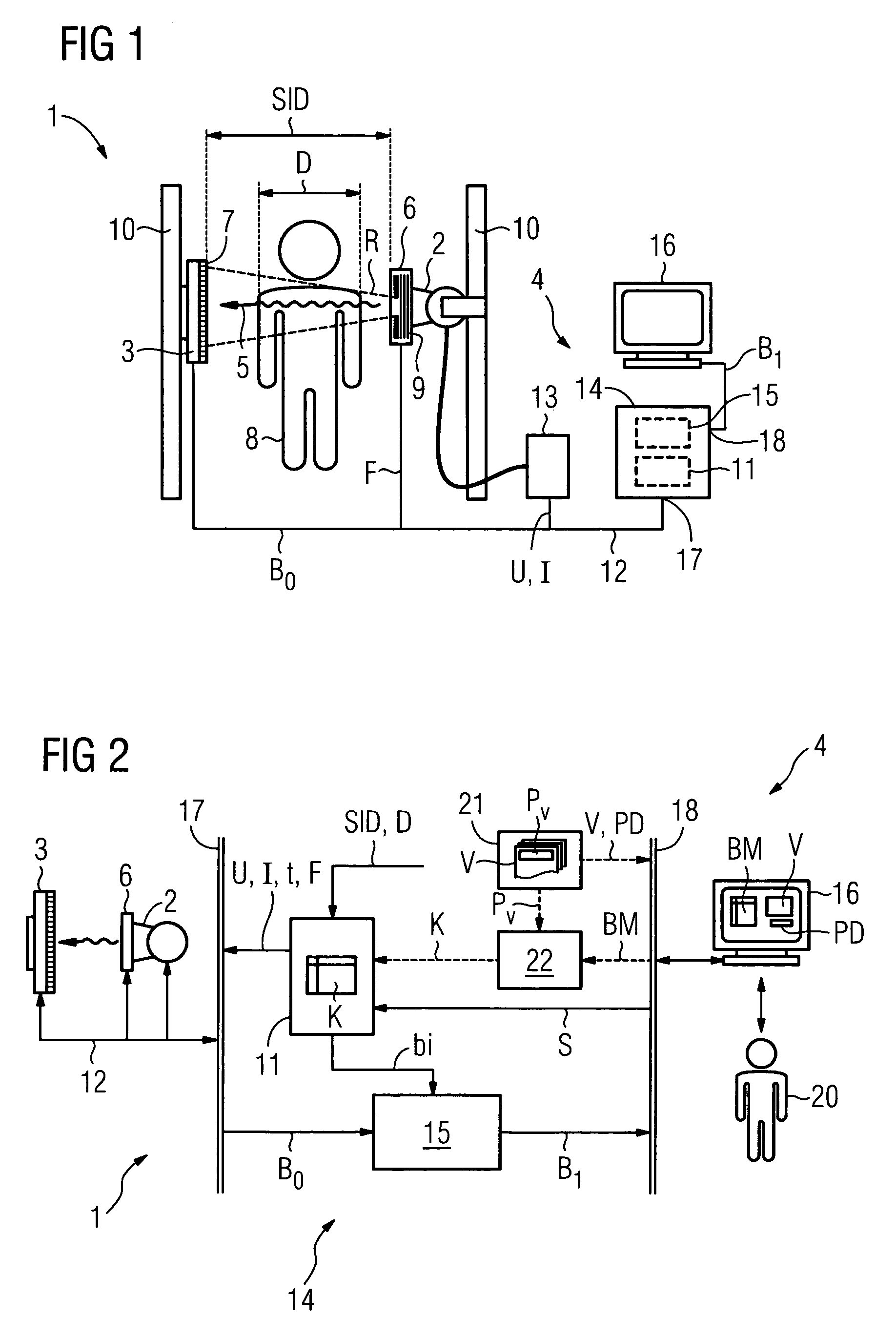
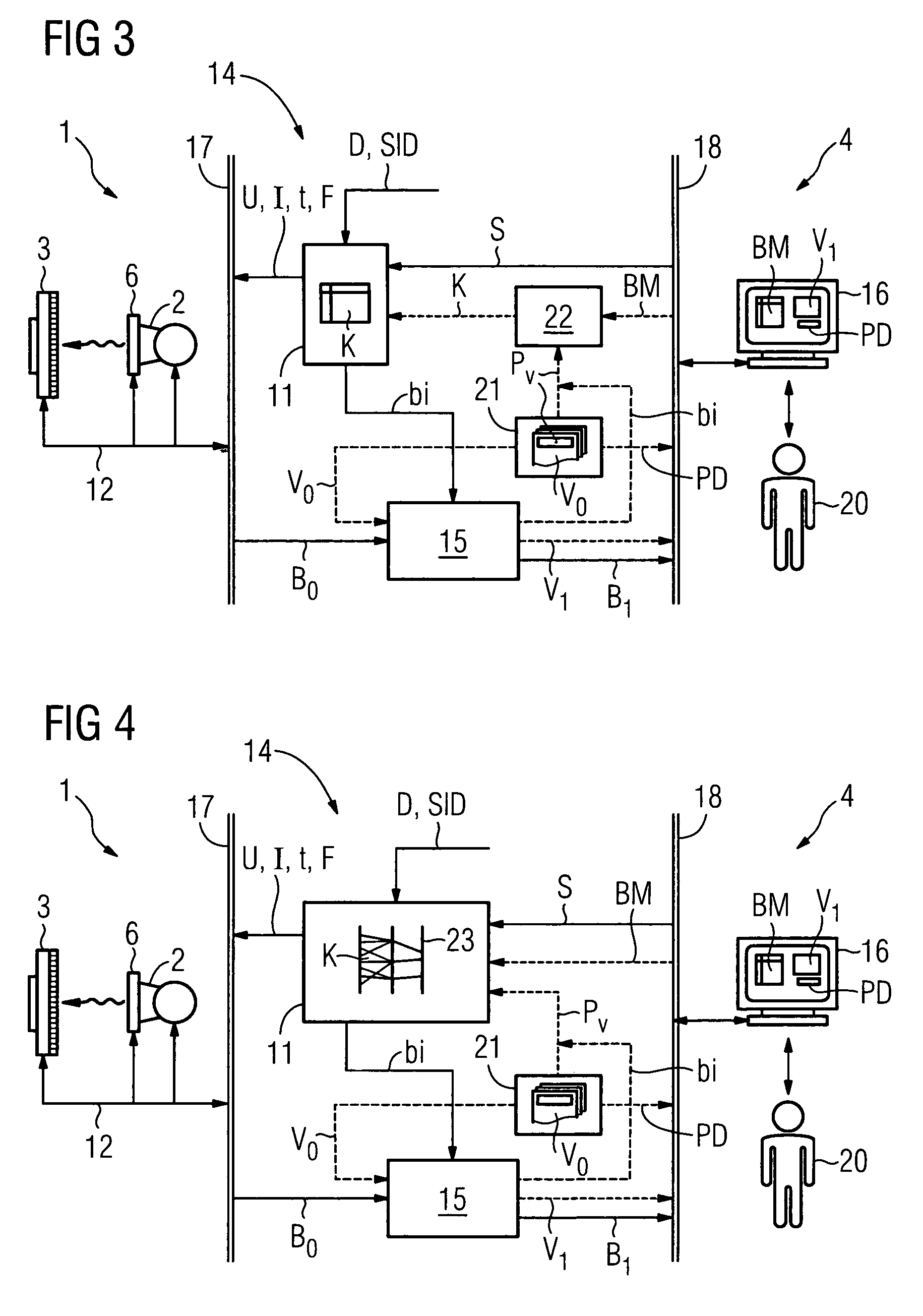
[0027]Mutually corresponding parts and variables are consistently denoted by the same reference characters in all the figures.

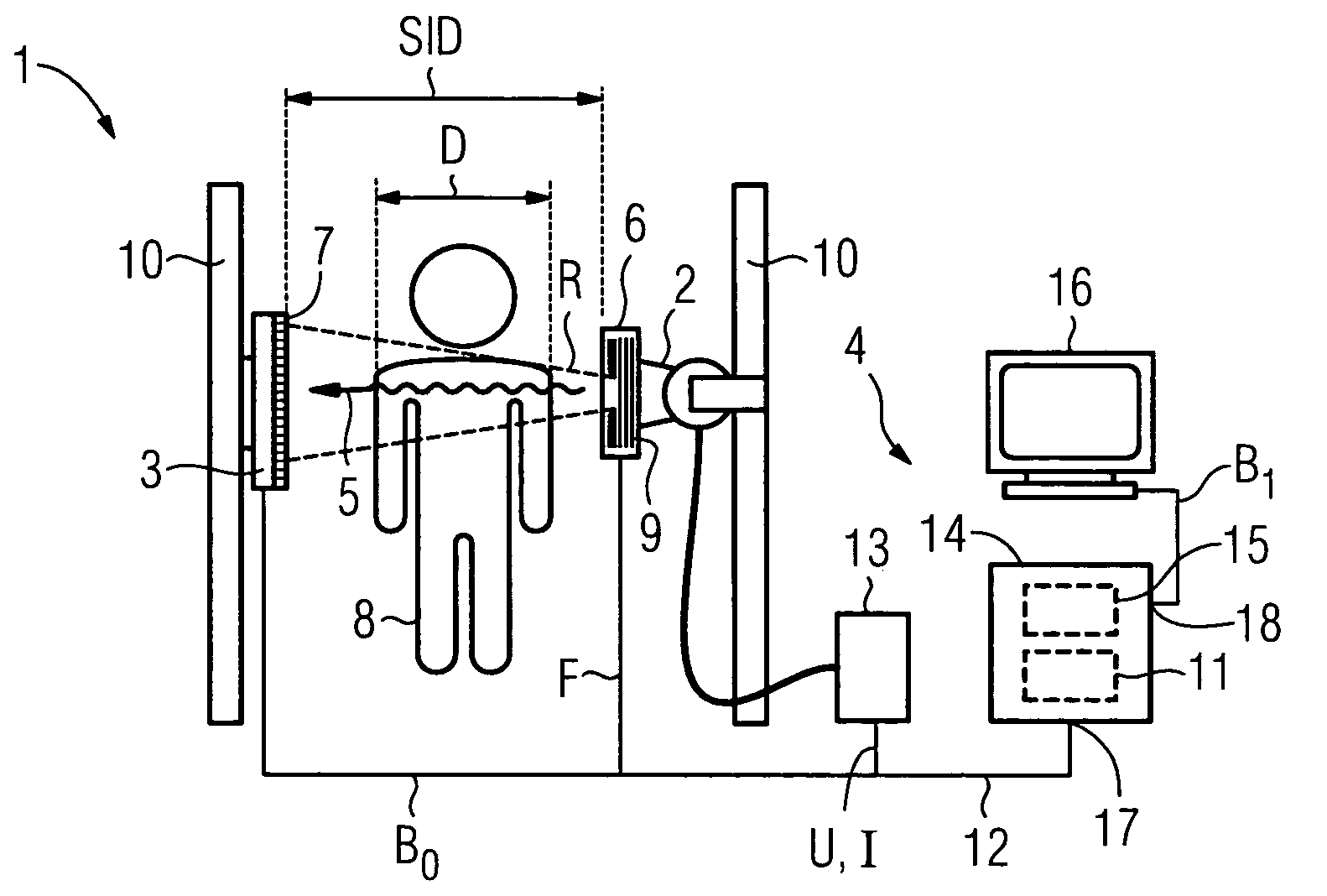
[0028]The medical x-ray device 1 schematically illustrated in FIG. 1 comprises an x-ray radiator 2, a digital x-ray detector 3 (hereinafter referred to as a detector for short) as well as a control and evaluation system 4. A collimator 6 and an anti-scatter grid 7 are interposed in a radiation direction 5 between the x-ray radiator 2 and the detector 3.

[0029]The collimator 6 is used to extract from the x-radiation R produced by the x-ray radiator 2 a sub-beam of a required size which passes through a patient under examination 8 or an object under examination and the anti-scatter grid 7 and is incident on the detector 3. The collimator 6 additionally contains a filter arrangement 9 by means of which the x-radiation R generated by the x-ray radiator 2 can be attenuated and / or modified in respect of its spectral distribution. The filter arrangement 9 is adjustab...


PUM


Abstract

A method and a suitable x-ray device (1) for carrying out the method are specified for effective and operationally simplified user-specific optimization of a parameter configuration (K) of an x-ray device (1), said configuration comprising at least one recording parameter (U,I,t,F). It is accordingly provided that a user (20) is shown a plurality of reference images (V,V1) for different reference parameter sets (PV) from a reference memory (21) in which are stored a large number of reference images (V,V0) each with an associated reference parameter set (PV), that for each reference image (V,V1) shown the user (20) submits an assessment (BM) of the image quality (V,V1), and that on the basis of the submitted assessments (BM) an optimized parameter configuration (K) is created from the reference parameter sets (PV) of the reference images (V,V1) shown.

Description

CROSS REFERENCE TO RELATED APPLICATIONS[0001]This application claims priority to the German application No. 10 2004 031 681.3, filed Jun. 30, 2004 which is incorporated by reference here in in its entirety.FIELD OF INVENTION[0002]The invention relates to a method for user-specific parameterization of an x-ray device designed in particular for angiographic and / or cardiological examinations according to the pre-characterizing clause of claim 1, and to an associated x-ray device according to the pre-characterizing clause of claim 8.BACKGROUND OF INVENTION[0003]For medical x-ray examinations in angiography and cardiology, excellent image quality is of particular importance in order to be able to differentiate clearly between the comparatively weakly absorbing structures being examined, in particular tissue and vessels as well as any catheters and stents present, in the body of a patient. At the same time, however, care must be taken to ensure that the patient and the medical personnel a...

Claims


Application Information

Patent Timeline
Patent Type & Authority Patents(United States)
IPC IPC(8): H05G1/30
CPCA61B6/4035A61B6/481A61B6/504A61B6/588A61B6/544A61B6/583A61B6/542
Inventor BERNHARDT, PHILIPPPFISTER, MARCUSSOLLACHER, RUDOLFTRESP, VOLKER
Owner SIEMENS HEALTHCARE GMBH
Why Eureka
Try Eureka