System and method for detecting a protrusion in a medical image

a protrusion and medical image technology, applied in image enhancement, tomography, instruments, etc., can solve the problems of many cad systems suffering from false positives, long analysis time, and high professional charges for radiologist interpretation

Inactive Publication Date: 2008-05-06
SIEMENS MEDICAL SOLUTIONS USA INC
View PDF3 Cites 11 Cited by
  • Summary
  • Abstract
  • Description
  • Claims
  • Application Information

AI Technical Summary

Problems solved by technology

However, a trained physician, clinician or radiologist may overlook a medical condition such as colonic polyps due to human error.
Although such CAD systems are very useful for diagnostic purposes, cost-reduction is difficult to achieve as the amount of data, for example, a radiologist, has to examine is abundant thus leading to lengthy analysis time and high costs of professional charges for the radiologist's interpretation.
In addition, many CAD systems suffer from false positives (e.g., incorrectly identifying normal tissues as abnormal) and false negatives (e.g., failing to correctly identify abnormalities).

Method used


Image

  • System and method for detecting a protrusion in a medical image
  • System and method for detecting a protrusion in a medical image
  • System and method for detecting a protrusion in a medical image

Examples


Embodiment Construction

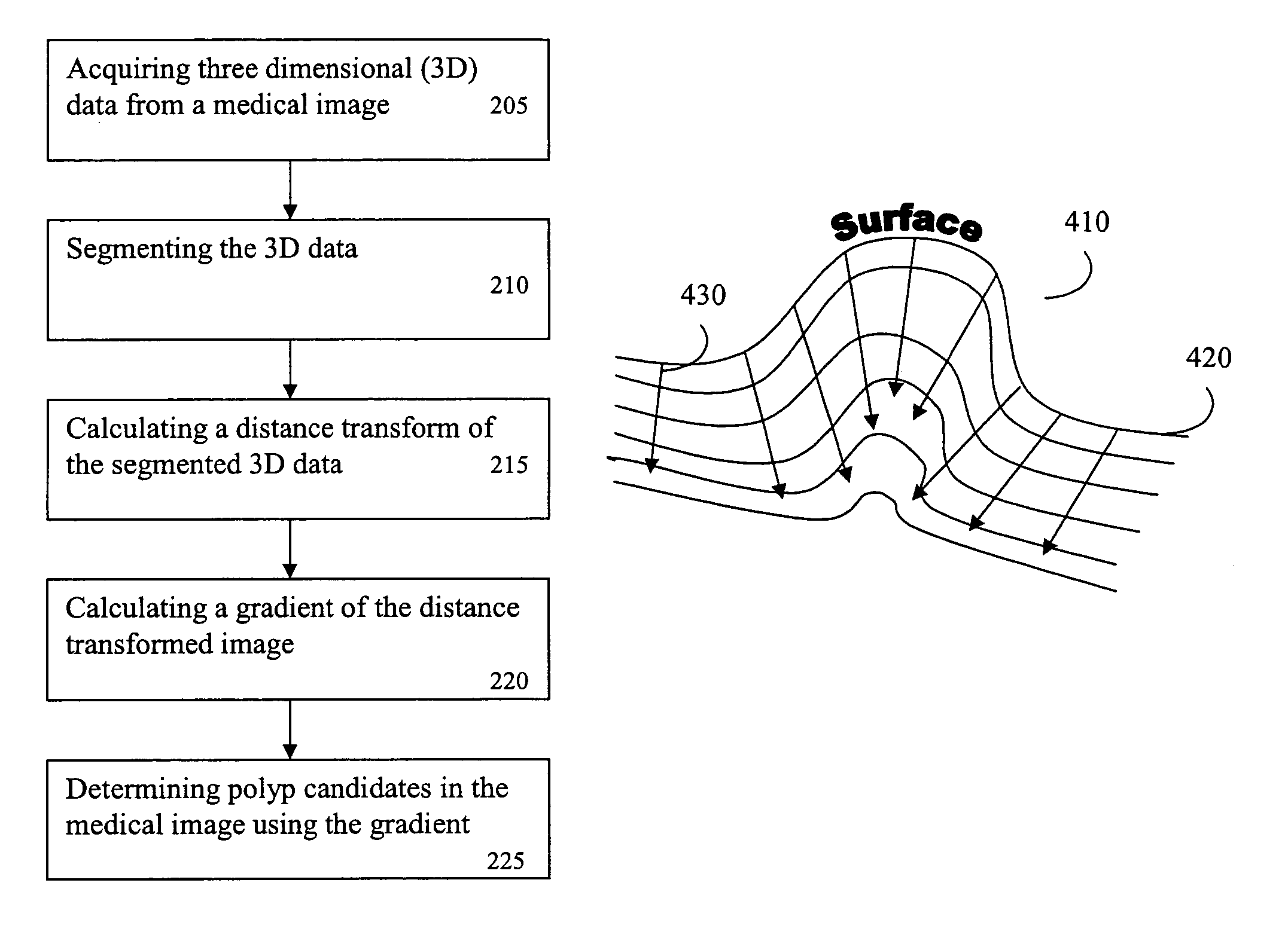
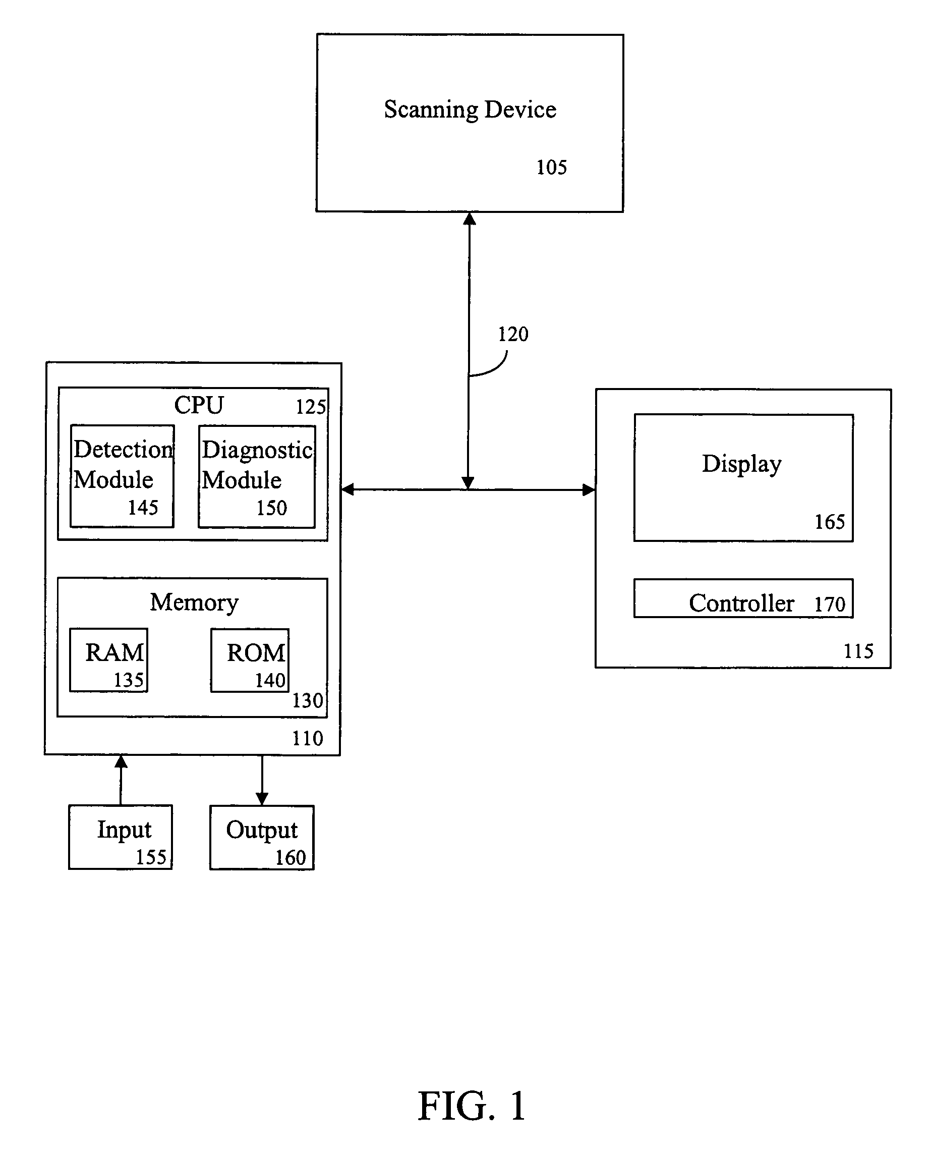
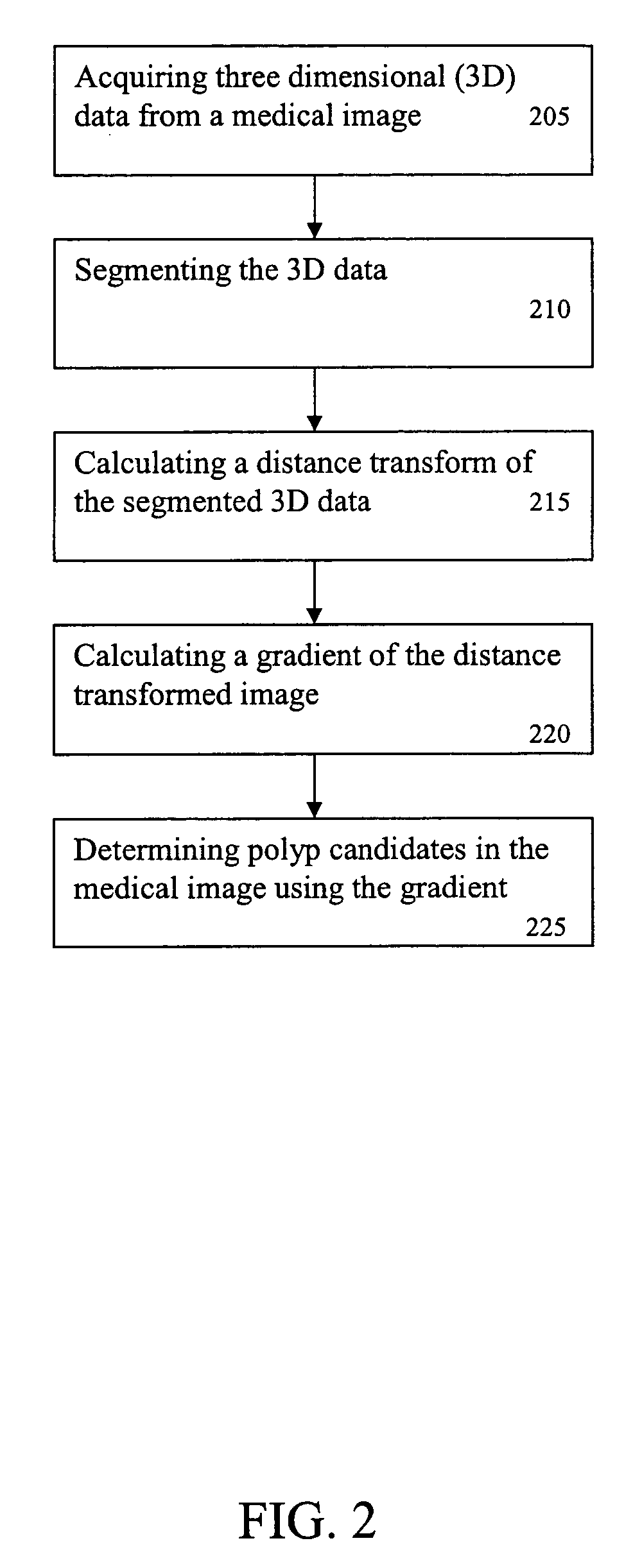
[0040]FIG. 1 is a block diagram of a system 100 for detecting a protrusion in a medical image according to an exemplary embodiment of the present invention. As shown in FIG. 1, the system 100 includes, inter alia, a scanning device 105, a personal computer (PC) 110 and an operator's console 115 connected over, for example, an Ethernet network 120. The scanning device 105 may be a magnetic resonance imaging (MRI) device, a computed tomography (CT) imaging device, a helical CT device, a positron emission tomography (PET) device, a two-dimensional (2D) or three-dimensional (3D) fluoroscopic imaging device, a 2D, 3D, or four-dimensional (4D) ultrasound imaging device, or an x-ray device, etc.

[0041]The PC 110, which may be a portable or laptop computer, a personal digital assistant (PDA), etc., includes a central processing unit (CPU) 125 and a memory 130, which are connected to an input 155 and an output 160. The CPU 125 includes a detection module 145, which is a computer-aided detecti...


PUM


Abstract

A method for detecting a protrusion in a medical image includes: acquiring a medical image, wherein the medical image is of an anatomical part; segmenting the medical image; calculating a distance map of the medical image; calculating a gradient of the distance mapped medical image; and processing the gradient to detect a protrusion in the medical image. The gradient is processed by: projecting a plurality of rays from a location in the distance mapped medical image; calculating a value for each of the plurality of rays based on features of each of the plurality of rays and the gradient of the distance mapped medical image; summing and scaling the value of each of the plurality of rays; and detecting one of a sphere-like and polyp-like shape using the summed and scaled values of the plurality of rays, wherein one of the sphere-like and polyp-like shapes is the protrusion.

Description

CROSS-REFERENCE TO RELATED APPLICATIONS[0001]This application claims the benefit of U.S. Provisional Application No. 60 / 486,799, filed Jul. 11, 2003, a copy of which is herein incorporated by reference.BACKGROUND OF THE INVENTION[0002]1. Technical Field[0003]The present invention relates to a system and method for detecting a protrusion in a medical image and, more particularly, to detecting a protrusion in a medical image by calculating a distance map of a segmented medical image and processing gradient characteristics of the distance mapped medical image to detect a protrusion in the medical image.[0004]2. Discussion of the Related Art[0005]In the field of medical imaging, various systems have been developed for generating medical images of various anatomical structures of individuals for the purposes of screening and evaluating medical conditions. These imaging systems include, for example, computed tomography (CT) imaging, magnetic resonance imaging (MRI), positron emission tomo...

Claims


Application Information

Patent Timeline
Patent Type & Authority Patents(United States)
IPC IPC(8): A61B6/03G06F19/00G06T7/00
CPCG06T7/0012H04N13/0022A61B6/03G06T2207/30028H04N13/128Y10S378/901
Inventor KIRALY, ATILLA PETERNOVAK, CAROL L.
Owner SIEMENS MEDICAL SOLUTIONS USA INC
Why Eureka
Try Eureka