
Enhanced transport using membrane disruptive agents

a technology of membrane disruption and enhanced transport, applied in the field of enhanced transport using membrane disruption agents, can solve problems such as membrane disruption, and achieve the effects of enhancing delivery, enhancing delivery or transport into or out of cells or through the skin

Inactive Publication Date: 2008-05-20
UNIV OF WASHINGTON +1
View PDF26 Cites 171 Cited by
  • Summary
  • Abstract
  • Description
  • Claims
  • Application Information

AI Technical Summary

Problems solved by technology

The lipid vehicles, especially cationic liposomes, may themselves cause membrane disruption.

Method used


Image

  • Enhanced transport using membrane disruptive agents
  • Enhanced transport using membrane disruptive agents
  • Enhanced transport using membrane disruptive agents

Examples


example 1

Evaluation of the pH sensitivity of PEAA-62K; PPAA

[0089]Objective:

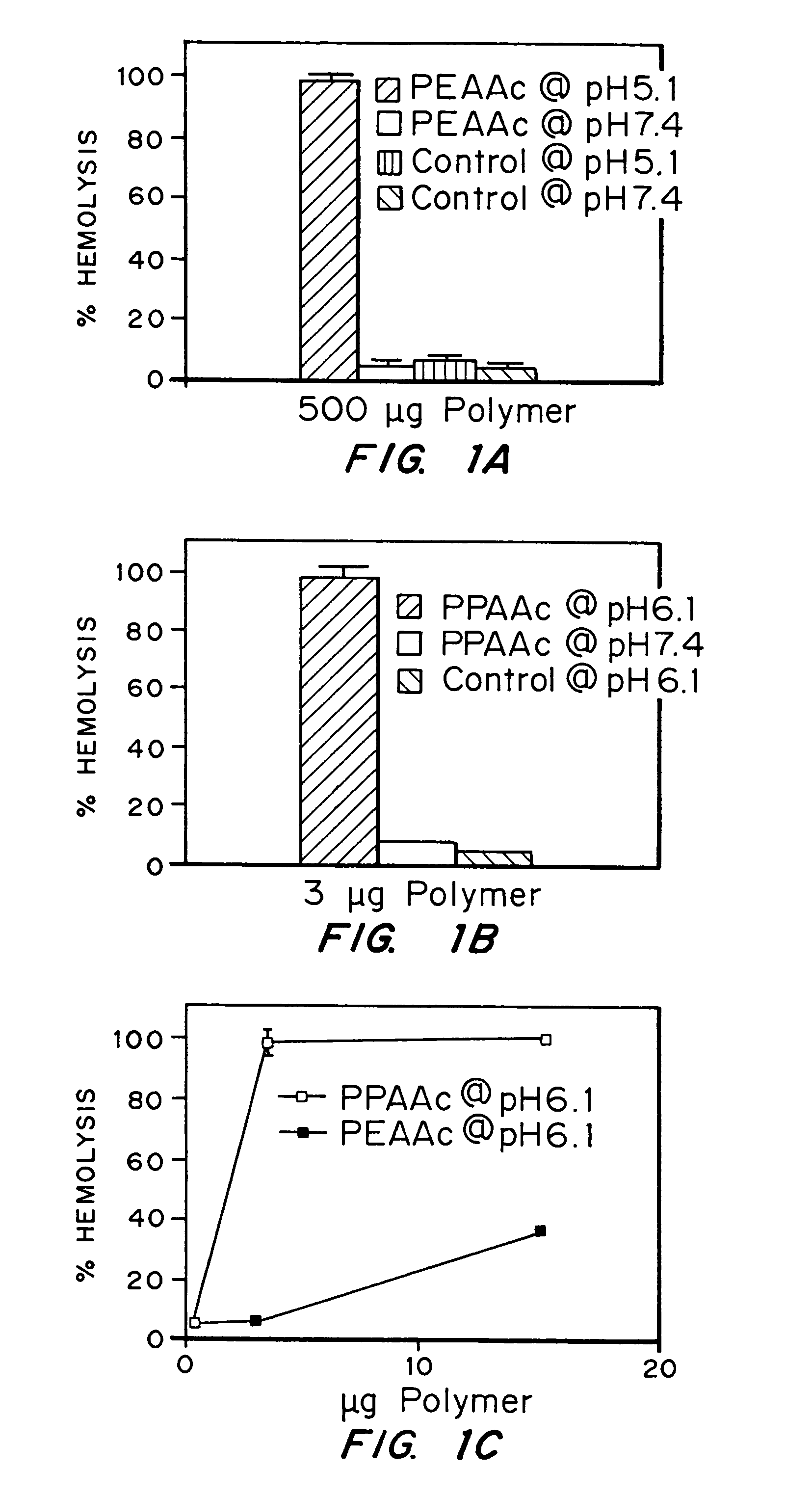
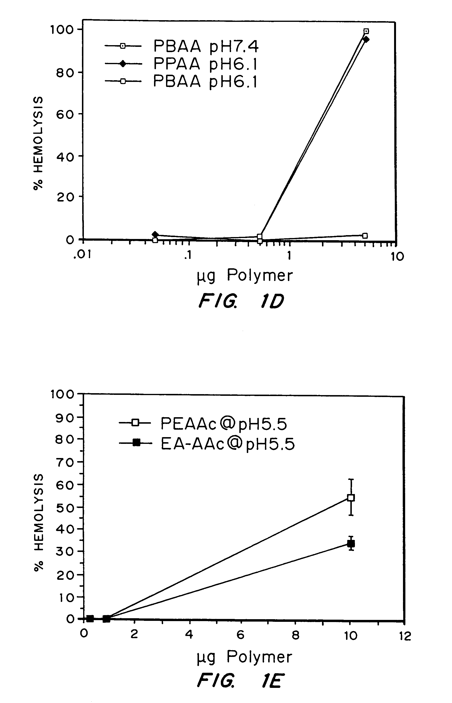
[0090]The objectives of this work were to determine if random copolymers of acrylic acid-butyl acrylate and acrylic acid-propyl acrylate have potential to act as endosomal releasing agents. This can be determined by measuring the hemolytic activity of the above polymers at endosomal pH (5.5) and physiologic pH (7.4).

[0091]Protocol:

[0092](I) Polymer Synthesis: Polymers and random copolymers of acrylic acid-propyl acrylate and acrylic acid-butyl acrylate were synthesized by free radical polymerization at various monomer feed ratios, in bulk, using AIBN as the initiator. The comonomer feed ratios are indicated in the relevant figures. The polymers were purified by ether precipitation.

(II) Hemolysis Assay: Fresh human blood was isolated in EDTA containing vacutainers, washed three times with 150 mM NaCl, and resuspended at a concentration of 108 cells / ml in PBS buffer at either pH 5.5 or pH 7.4 or MES buffer, as noted. Th...

example 2

Comparison of the pH Sensitivity of EALA with that of an EALA / Polyacrylic Acid Conjugate

[0106]The ability of EALA to lyse erythrocytes was compared with that of an EALA / polyacrylic acid conjugate at a pH of 5.0 using approximately 107 red blood cells in 100 mM dibasic NaPO4, incubated at 37° C. for 20 minutes. A physical mixture of EALA and PAA was also tested.

[0107]The results are shown in FIG. 2. The EALA peptide by itself, as well as the physical mixture of EALA with PAA, demonstrated a negligible amount of lysis whereas the conjugate yielded about 70% lysis at a concentration of about 100 μg.

example 7

Comparison of the pH Sensitivity of PEAA with that of an IgG / PEAA Conjugate and that of IgG Alone

[0108]The ability of PEAA, an IgG / PEAA conjugate, and IgG alone to lyse erythrocytes was compared by performing a hemolysis assay at a pH of 5.0 using approximately 108 red blood cells in 100 mM dibasic sodium phosphate, and incubating at 37° C. for an hour. 1-ethyl-3-(3-dimethylaminopropyl)carbodiimide hydrochloride (EDC) was used to conjugate PEAA to rabbit immunoglobulin G (IgG). EDC reacts with the carboxyl groups to PEAA to form an amine-reactive intermediate, which then reacts with the lysine amine groups on IgG. The IgG was oxidized using 100 mM sodium periodate to yield reactive aldehyde groups on the carbohydrate moiety. Conjugation was realized via Schiff base formation between the end amine group of the amine-terminated PEAA and the aldehyde group on the IgG. This bond is reduced using 5 M sodium cyanoborohydride to yield a covalently bound conjugate of PEAA and IgG. The molar...


PUM

PropertyMeasurementUnit
pHaaaaaaaaaa
pHaaaaaaaaaa
pHaaaaaaaaaa

Abstract

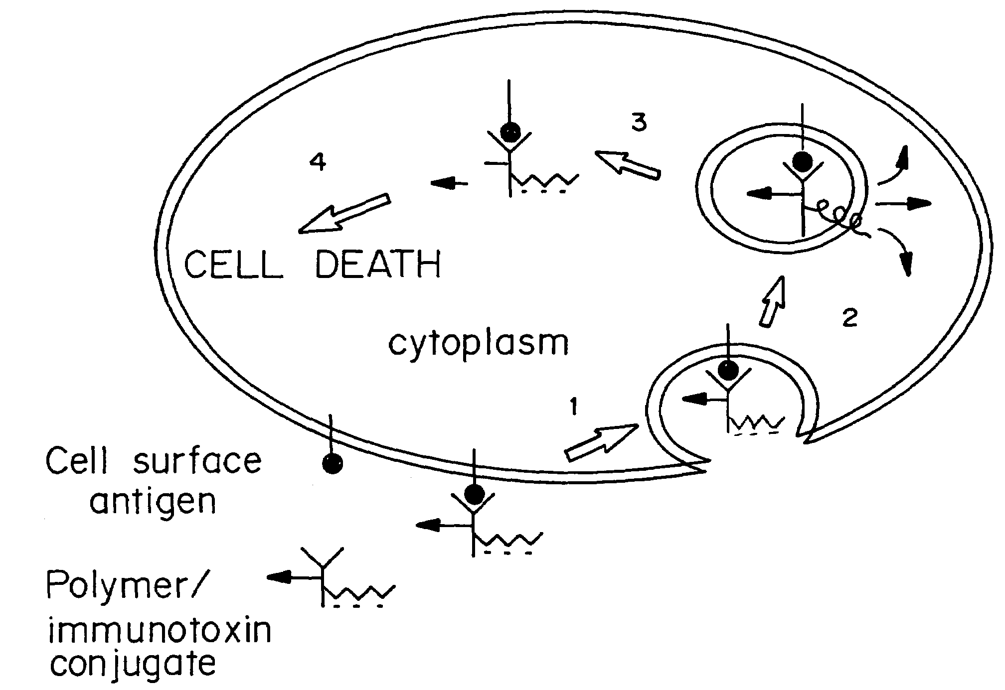
Compositions and methods for transport or release of therapeutic and diagnostic agents or metabolites or other analytes from cells, compartments within cells, or through cell layers or barriers are described. The compositions include a membrane barrier transport enhancing agent and are usually administered in combination with an enhancer and / or exposure to stimuli to effect disruption or altered permeability, transport or release. In a preferred embodiment, the compositions include compounds which disrupt endosomal membranes in response to the low pH in the endosomes but which are relatively inactive toward cell membranes, coupled directly or indirectly to a therapeutic or diagnostic agent. Other disruptive agents can also be used, responsive to stimuli and / or enhancers other than pH, such as light, electrical stimuli, electromagnetic stimuli, ultrasound, temperature, or combinations thereof. The compounds can be coupled by ionic, covalent or H bonds to an agent to be delivered or to a ligand which forms a complex with the agent to be delivered. Agents to be delivered can be therapeutic and / or diagnostic agents. Treatments which enhance delivery such as ultrasound, iontopheresis, and / or electrophereis can also be used with the disrupting agents.

Description

CROSS-REFERENCES TO RELATED APPLICATIONS[0001]This application is a continuation of U.S. patent application Ser. No. 09 / 226,044, filed Jan. 5, 1999 now U.S. Pat. No. 6,835,393, which claims the benefit of U.S. Provisional Application No. 60 / 070,411, filed Jan. 5, 1998. The above-referenced applications are incorporated herein by reference in their entirety.STATEMENT REGARDING FEDERALLY FUNDED RESEARCH[0002]The U.S. government has certain rights in this invention by virtue of an National Institutes of Health grant, National Institutes General Medical Sciences grant GM 53771-02.[0003]The present invention is in the field of the delivery of therapeutic agents, and more particularly in the area of enhancement of transport or delivery of molecules into the cell cytosol, through cell barriers or layers of cells, or through lipid membranes, using membrane barrier transport enhancing agents alone or in combination with a stimulus and / or enhancer that modifies the structure and / or properties...

Claims


Application Information

Patent Timeline
Patent Type & Authority Patents(United States)
IPC IPC(8): A61K9/127A61K47/00A61K9/14A61K9/16A61K47/32A61K9/00A61K31/7105A61K31/711A61K41/00A61K47/42A61K47/48
CPCA61K41/0028A61K41/0047A61K47/48176A61K47/481A61K47/48376A61K47/6801
Inventor HOFFMAN, ALLAN S.STAYTON, PATRICKPRESS, OLIVER W.MURTHY, NIRENREED, CHANTAL LACKEYCRUM, LAWRENCE A.MOURAD, PIERRE D.PORTER, TYRONE M.TIRRELL, DAVID
Owner UNIV OF WASHINGTON
Learn More