System and method for segmenting the left ventricle in a cardiac image

a technology of cardiac image and left ventricle, applied in the field of medical imaging, can solve the problems of not having a known system or method for providing an adaptive technique for analyzing cardiac image segmentation, and the difficulty of segmentation of these images

Inactive Publication Date: 2008-07-15
SIEMENS MEDICAL SOLUTIONS USA INC
View PDF10 Cites 11 Cited by
  • Summary
  • Abstract
  • Description
  • Claims
  • Application Information

AI Technical Summary

Problems solved by technology

Segmentation of these images has been particularly challenging.
However, no known system or method exists for providing an adaptive technique of analyzing cardiac images.

Method used


Image

  • System and method for segmenting the left ventricle in a cardiac image
  • System and method for segmenting the left ventricle in a cardiac image
  • System and method for segmenting the left ventricle in a cardiac image

Examples


Embodiment Construction

[0038]A system and / or method for segmenting a cardiac image of a left ventricle can be embodied in any suitable commercial cardiac analysis package, such as the ARGUS cardiac analysis package from Siemens, which offers a complete system of drawing tools and automatic segmentation methods to allow the physician to outline the myocardium in each image in the patient data set, determine volumes, ejection fraction, and perform a thickening analysis.

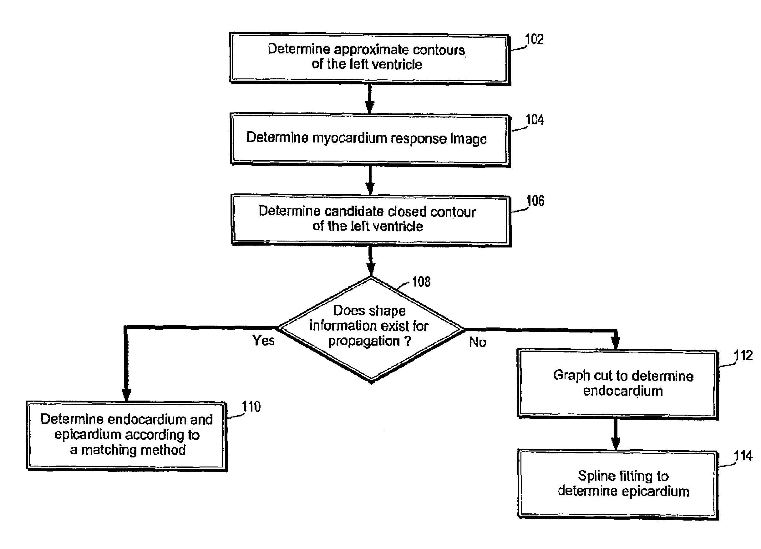
[0039]Referring to FIG. 1, illustrating a method according to an embodiment of the present disclosure, the method determines approximate contours of the left ventricle 102 and a myocardium response image 104 according to a histogram of pixel intensity in the magnetic resonance image of interest. The method determines a plurality of candidate closed contours of the left ventricle 106, according to a plurality of energy functions. Further, the method is aware of whether shape information exists for the magnetic resonance image of interest 108 a...


PUM


Abstract

A method is provided for segmenting an image of interest of a left ventricle. The method includes determining a myocardium contour according to a graph cut of candidate endocardium contours, and a spline fitting to candidate epicardium contours in the absence of shape propagation. The method further includes applying a plurality of shape constraints to candidate endocardium contours and candidate epicardium contours to determine the myocardium contour, wherein a template is determined by shape propagation of a plurality of images in a sequence including the image of interest in the presence of shape propagation.

Description

CROSS-REFERENCE TO RELATED APPLICATIONS[0001]This is a continuation-in-part application of U.S. application Ser. No. 09 / 970,552, filed Oct. 4, 2001, now U.S. Pat. No. 6,961,454 entitled “System and Method for Segmenting the Left Ventricle in a Cardiac MR Image”, the disclosure of which is herein incorporated by reference in its entirety.BACKGROUND OF THE INVENTION[0002]1. Technical Field[0003]The present disclosure relates to medical imaging, and more particularly to extracting the myocardium from four-dimensional image data (two-dimensional images over time and space).[0004]2. Discussion of Related Art[0005]Cardiovascular disease is the leading cause of death in the United States. Mortality has been declining over the years as lifestyle has changed, but the decline is also due to the development of new technologies to diagnose disease. One of these techniques is magnetic resonance imaging (MRI), which provides time-varying three-dimensional imagery of the heart. To help in the diag...

Claims


Application Information

Patent Timeline
Patent Type & Authority Patents(United States)
IPC IPC(8): G06K9/00A61B5/055G06T5/00G06V10/26
CPCA61B5/055G06K9/34G06T7/0012G06T7/0083G06T7/0093G06T2207/30048G06T2207/10081G06T2207/10088G06T2207/20061G06T2207/20164A61B2576/023G06T7/12G06T7/162G16H30/40G06V10/26
Inventor JOLLY, MARIE-PIERRESUN, YINGDUTA, NICOLAE
Owner SIEMENS MEDICAL SOLUTIONS USA INC
Why Eureka
Try Eureka