Apparatus and method for removing abnormal tissue

a technology of abnormal tissue and apparatus, applied in the field of surgical equipment and methods, can solve the problems of limited biopsy method, limited excision of abnormality, limited biopsy method, etc., and achieve the effects of reducing scarring and deformation of remaining tissue, limiting the volume of tissue removed, and eliminating cost and risk

Inactive Publication Date: 2008-10-21
CHAUHAN SUNITA +3
View PDF12 Cites 137 Cited by
  • Summary
  • Abstract
  • Description
  • Claims
  • Application Information

AI Technical Summary

Benefits of technology

[0013]Methods exemplary of the invention use the apparatus to provide an assisted or automated surgical method for removing abnormal tissue, such as a tumour, from a breast, and which limit the volume of tissue removed and the size of the incision or incisions made, so as to reduce scarring and deformation of the remaining tissue. The method is particularly suitable for small breast tumours and abnormalities that are localized and intact (e.g. carcinomas in situ). It can be performed using a local anaesthetic on an outpatient basis, eliminating the cost and risk associated with current lumpectomy and mastectomy procedures.

Problems solved by technology

The existing biopsy methods are limited to one or at most a few sample removals, and are not designed, nor are intended, to perform surgical excision of the abnormality.
In some cases the biopsy method is limited to the removal of just enough cells or fluid sufficient for pathological testing.
Surgical methods typically require stitches to close the excision and can leave a scar, which may complicate the interpretation of follow up breast examinations; increase the chance of bleeding, and infection; and cause problems with wound healing.
As well, there are mortality risks associated with the use of anaesthesia.

Method used


Image

  • Apparatus and method for removing abnormal tissue
  • Apparatus and method for removing abnormal tissue
  • Apparatus and method for removing abnormal tissue

Examples


Embodiment Construction

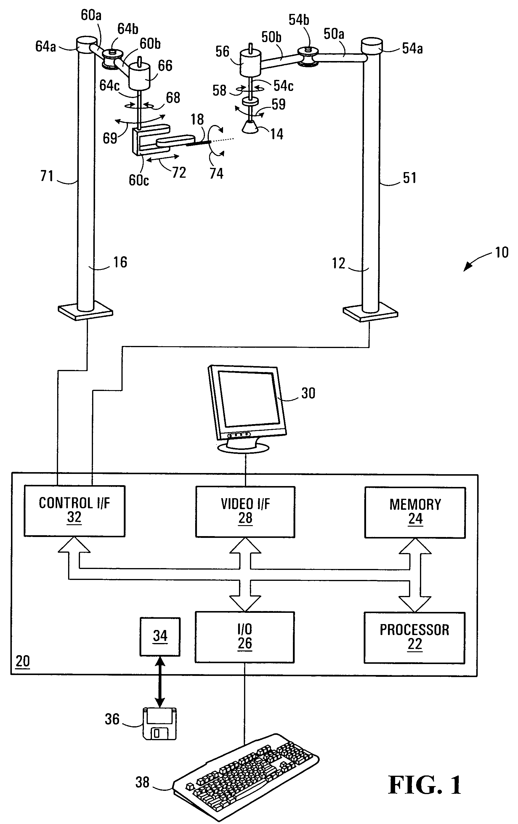
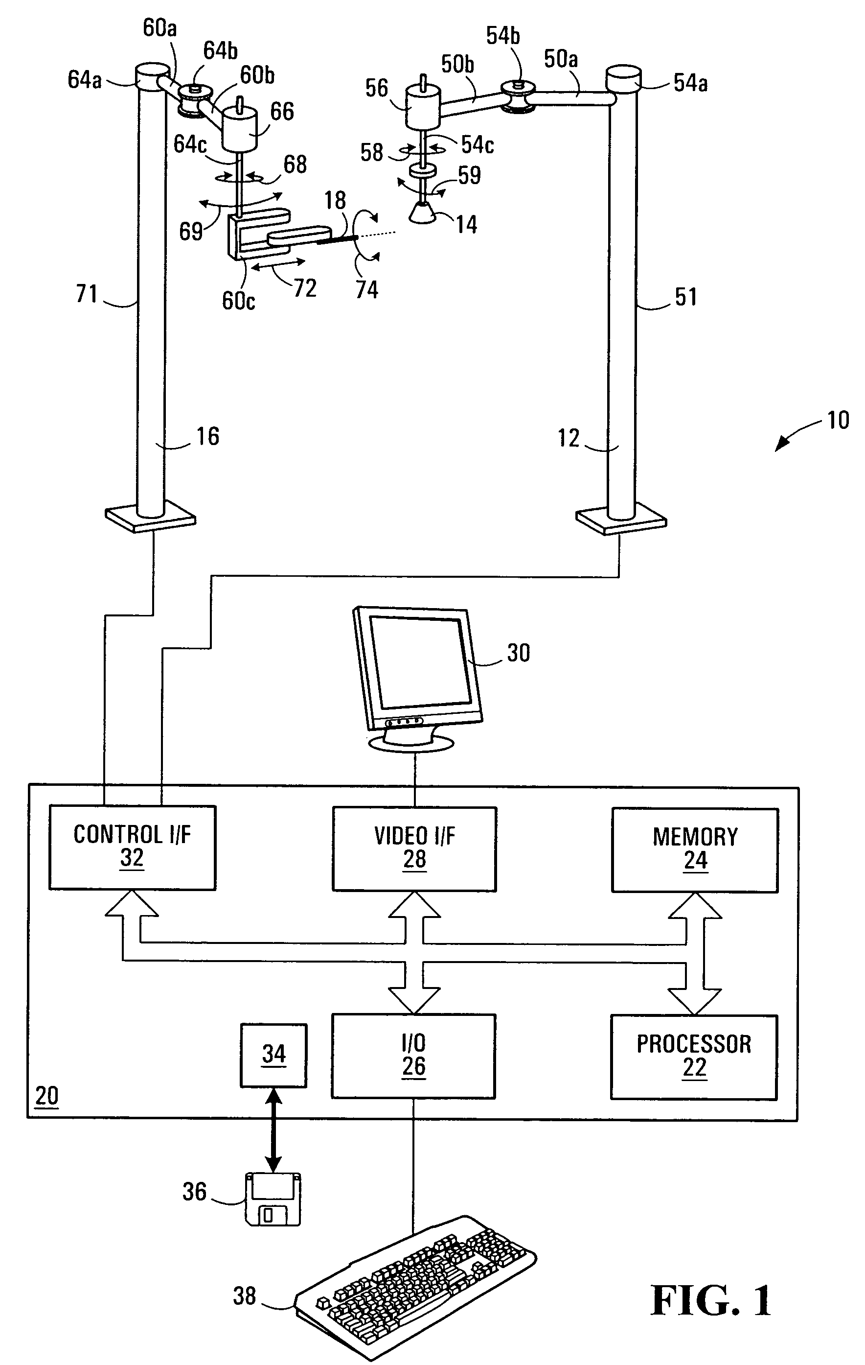
[0032]FIG. 1 illustrates an apparatus 10 for removing abnormal tissue from the breast of a patient, exemplary of an embodiment of the present invention.

[0033]In this context, the term “abnormal tissue” refers to tissue, which may be benign or malignant, and includes cysts, lumps, microcalcifications, spiculated masses, and asymmetric densities, and which may be palpable or non-palpable. Other abnormal tissue may be part of localized tumors, including carcinoma—in situ, such as Fibroadenomas, Phyllodes tumours, which are benign breast tumors in the glandular and stroma (connective), Fat necrosis (a benign condition where fatty breast tissue swells or becomes tender) as well as malignant breast tumours such as Lobular Carcinoma in situ (LCIS), Ductal Carcinoma in situ (DCIS), preferably at Stage-0 and I.

[0034]As illustrated, apparatus 10 includes a robotic imaging arm 12 for holding and moving an imaging probe 14; and a robotic surgical arm 16 for holding and moving a minimally invasi...


PUM


Abstract

A computer assisted, minimally invasive method and apparatus for surgically removing abnormal tissue from a patient, for example, from a breast, are disclosed. The method involves imaging of the breast to locate the abnormal tissue, and determining a volume encapsulating the abnormal tissue and including a margin of healthy tissue. Based on the volume, a sequence of movements of a surgical instrument for tissue removal device is planned, so as to predictably excise the desired volume of tissue.

Description

FIELD OF THE INVENTION[0001]The present invention relates generally surgical apparatus and methods, and more particularly to an apparatus and method for removing abnormal tissue. The apparatus and methods are particularly well suited for removing abnormal breast tissue.BACKGROUND OF THE INVENTION[0002]In most countries, breast cancer is the second most prevalent cancer to afflict women. Currently, abnormal breast tissue is detected by various methods, such as physical breast examination, mammography, ultrasound and other breast imaging methods.[0003]In order to determine whether such abnormalities are benign or malignant, examination of the histopathology of the abnormal tissue is required. Therefore, a sample of the abnormal tissue is taken by biopsy and is analysed by microscopic evaluation.[0004]At present, there are biopsy techniques that can be performed through a small incision in the affected breast, and which are therefore considered to be minimally invasive. Typically, a ra...

Claims


Application Information

Patent Timeline
Patent Type & Authority Patents(United States)
IPC IPC(8): A61B5/05A61B8/00A61N1/30
CPCA61B19/2203A61B19/52A61B19/201A61B19/26A61B19/50A61B2017/008A61B2019/2234A61B90/36A61B90/50A61B34/30A61B90/11A61B2034/305A61B34/10
Inventor CHAUHAN, SUNITAMISHRA, RANJAN KUMARWEE, SIEW BOCKTAN, MONA P.
Owner CHAUHAN SUNITA
Why Eureka
Try Eureka