Method and apparatus for detecting endometriosis

Inactive Publication Date: 2008-12-09
JARRELL JOHN F
View PDF10 Cites 10 Cited by
  • Summary
  • Abstract
  • Description
  • Claims
  • Application Information

AI Technical Summary

Benefits of technology

[0060]As this method is non-surgical in nature, a number of diagnostic treatments using the present invention can be made on a patient without the high costs and limited access of MRI or the dangers or risks caused by multiple laparotomy or laparoscopy procedures. Accordingly, the present invention can be used as part of regular examinations to detect the early onset of endometriosis in a patient. The present invention can also be used at regular intervals while the patient is undergoing medical treatment for endometriosis to determine the efficacy of the treatment.

Problems solved by technology

The amounts of iron appear to correlate roughly with the extent of endometriosis although this is empiric and has not been subjected to a specific analysis owing to the difficulties associated with such research.

Method used


Image

  • Method and apparatus for detecting endometriosis
  • Method and apparatus for detecting endometriosis
  • Method and apparatus for detecting endometriosis

Examples


Embodiment Construction

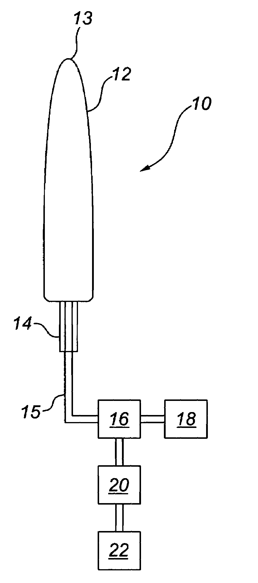
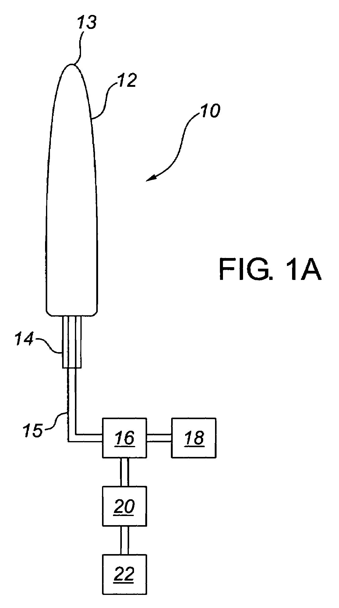
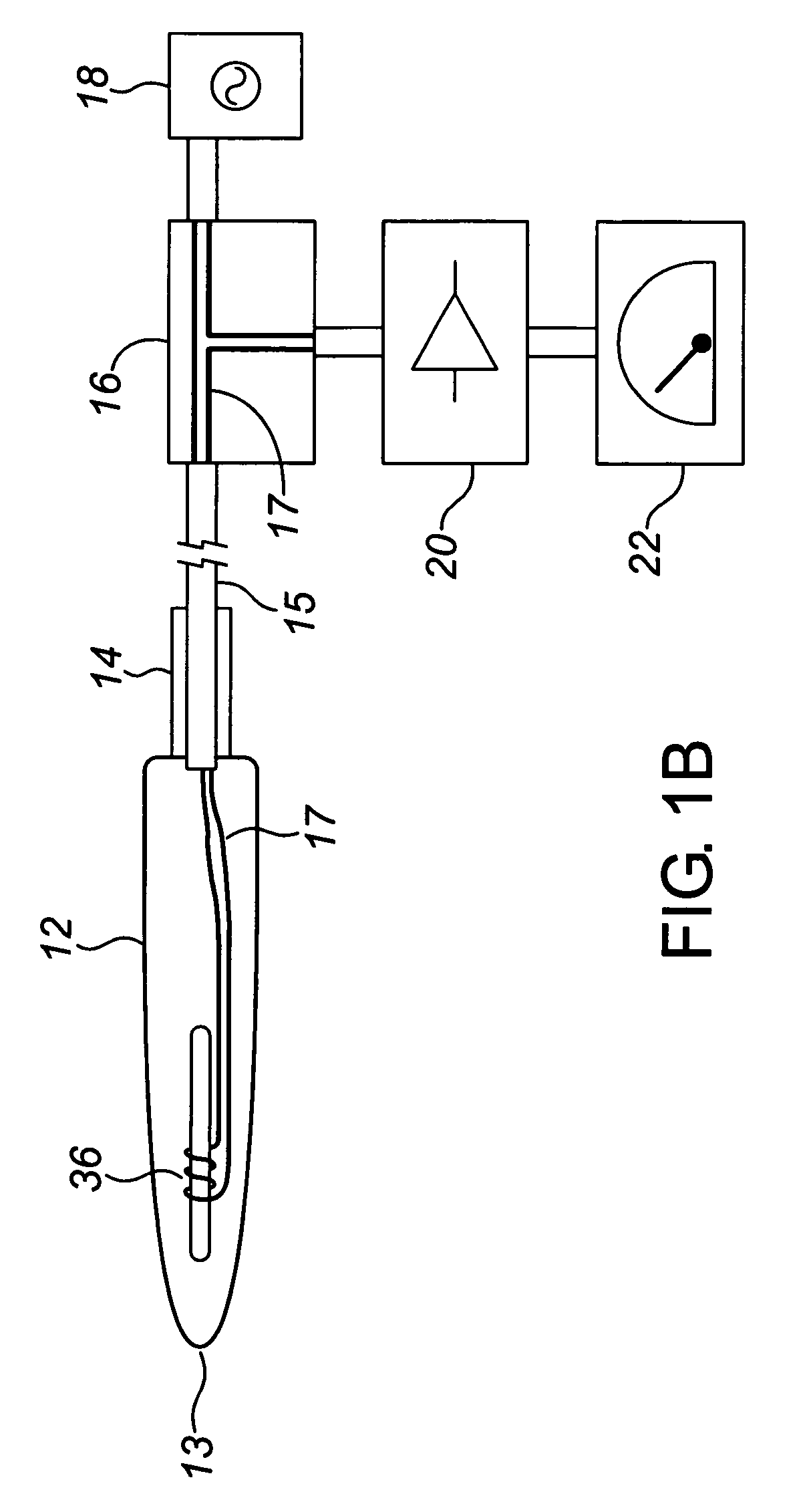
[0071]The present invention is a method and apparatus for detecting endometriosis. According to FIGS. 1A and 1B, one embodiment of the present invention is shown which comprises of Apparatus 10. Apparatus 10 comprises a probe 12 with handle 14. Probe 12 is connected to balancing unit 16 via cable 15. Bias current for coil 36 is provided by excitation source 18 through balancing unit 16 and wires 17. Balancing unit 16 is also connected to amplifier 20 which, in turn, is connected to indicator 22.

[0072]When probe 12 is brought in proximity of metal or ferrous material, such as endometriotic tissue, the current in coil 36 fluctuates due to the presence of metal near coil 36. Amplifier 20 amplifies fluctuations in the current. The amplified fluctuations are then displayed on indicator 22. Indicator 22 may be an analog or digital display giving an indicator of current or voltage as a representation of the bias current fluctuations. Apparatus 10 may be configured to provide qualitative re...


PUM

PropertyMeasurementUnit
magnetic fieldaaaaaaaaaa
timeaaaaaaaaaa
MRIaaaaaaaaaa

Abstract

A vaginal probe includes a metal detection sensor for detecting the presence of ferrous-laden endometriotic tissues. The probe is electrically connected to external electronic circuitry for interpreting the readings taken by the sensor and for providing a qualitative or quantitative measurement of the amount of endometriotic tissues present. The probe is inserted in either the vagina or peritoneal cavity of a patient to detect the location and the relative amounts of iron or ferrous-laden tissues in the pelvic area of the patient. Using the probe and monitoring the readings over time as part of pelvic exams assists in determining the early onset or progression of endometriosis and / or the response to one or more therapies being provided to the patient to treat the disease.

Description

CROSS REFERENCE TO RELATED APPLICATIONS[0001]This application claims priority from U.S. Provisional Application No. 60 / 553,055 filed Mar. 15, 2004, entitled “Method and Apparatus for Detecting Endometriosis,” which is incorporated herein by reference.BACKGROUND OF THE INVENTION[0002]The present invention relates to the field of diagnostic methods and apparatus for detecting endometriosis in a patient. More specifically, the present invention relates to a non-surgical method and apparatus for detecting endometriosis.[0003]Endometriosis is defined as the presence of endometrial tissue in aberrant locations of the body, usually in the pelvis.[0004]There are no techniques for the confirmation of this disease other than the pathological assessment of the tissue that is removed or biopsied during a surgical procedure. Magnetic Resonance Imaging (“MRI”) is helpful in the detection but the standard of care still requires histological confirmation. The pathological definition is based on the...

Claims


Application Information

Patent Timeline
Patent Type & Authority Patents(United States)
IPC IPC(8): A61B5/00A61B5/04A61B5/05A61B10/00G01N33/48
CPCA61B5/05A61B5/00A61B10/00G01N33/48
Inventor JARRELL, JOHN F.
Owner JARRELL JOHN F
Why Eureka
Try Eureka