Method and device for percutaneous surgical ventricular repair

a technology of ventricular repair and percutaneous surgery, applied in the field of percutaneous surgical ventricular repair, can solve the problems of no longer being able to contribute to the squeezing or twisting motion required to pump blood, ischemic muscle is no longer capable of contracting, and blood pressure tends to bulge or expand the chamber

Inactive Publication Date: 2009-02-03
HEART FAILURE TECH
View PDF143 Cites 83 Cited by
  • Summary
  • Abstract
  • Description
  • Claims
  • Application Information

AI Technical Summary

Benefits of technology

[0017]In response to these and other problems, an improved apparatus and method is provided for endoscopic surgical ventricle repair which allows a surgeon to perform a surgical ventricular repair procedure through a closed chest or through a small thoracotomy on a beating, fibrillating or an arrested heart. In one embodiment, there is a method for repairing a heart of a human, comprising introducing a shaping device percuta

Problems solved by technology

The ischemic muscle is no longer capable of contracting so it cannot contribute to either squeezing or twisting motion required to pump blood.
In severe cases the akinetic tissue, which is not capable of contracting, is in fact elastic so that blood pressure tends to develop a bulge or expansion of the chamber.
This is particularly detrimental to the limited pumping action available, as the heart loses even more of its energy to pumping the bulge instead of the blood.
As a consequence, the fibers are poorly oriented to accomplish even the squeezing action, as the lines of force become less perpendicular to the heart wall.
The spherical shape further reduces pumping efficiency since the fibers which normally encircle the apex facili

Method used


Image

  • Method and device for percutaneous surgical ventricular repair
  • Method and device for percutaneous surgical ventricular repair
  • Method and device for percutaneous surgical ventricular repair

Examples


Embodiment Construction

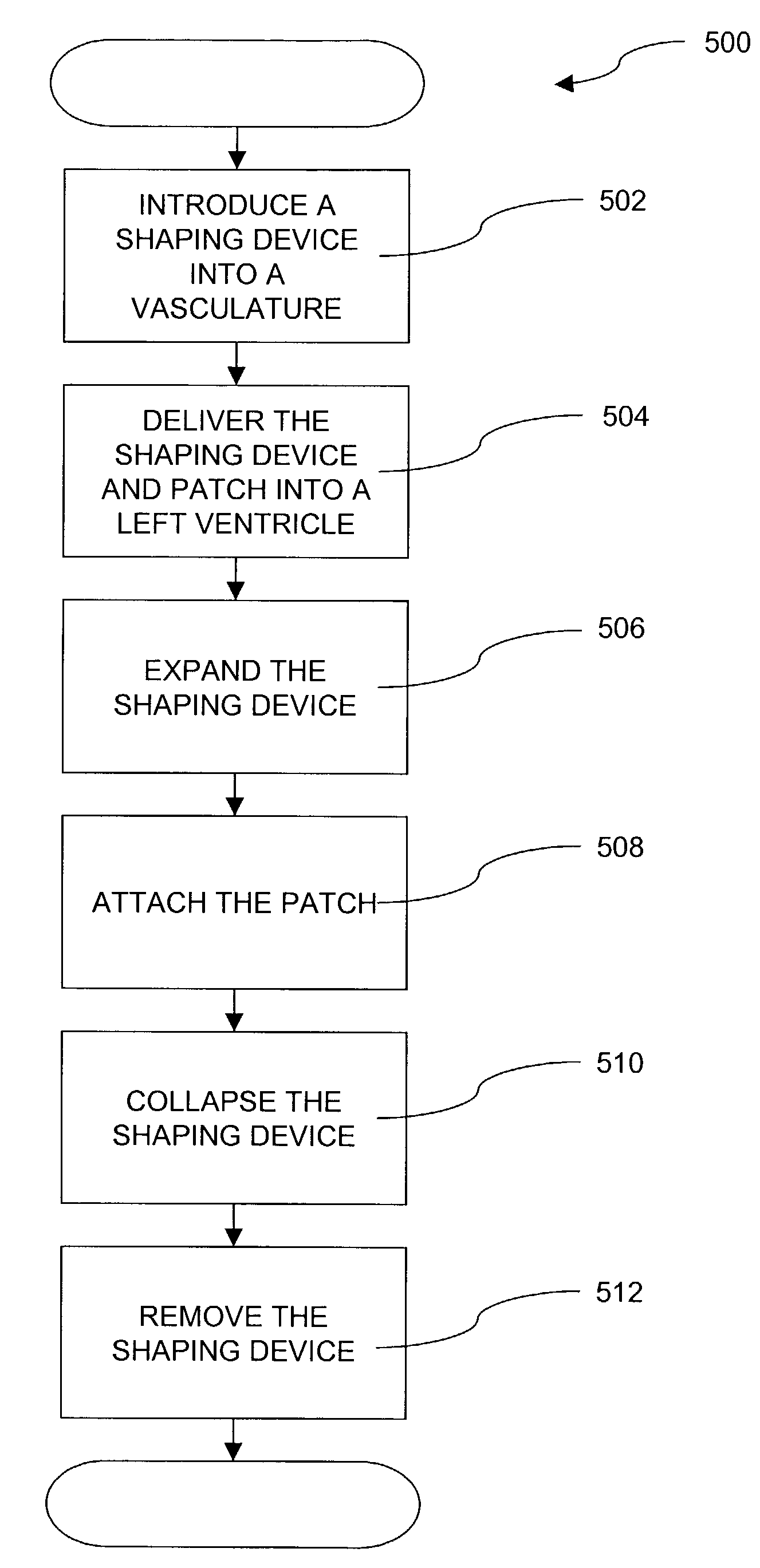
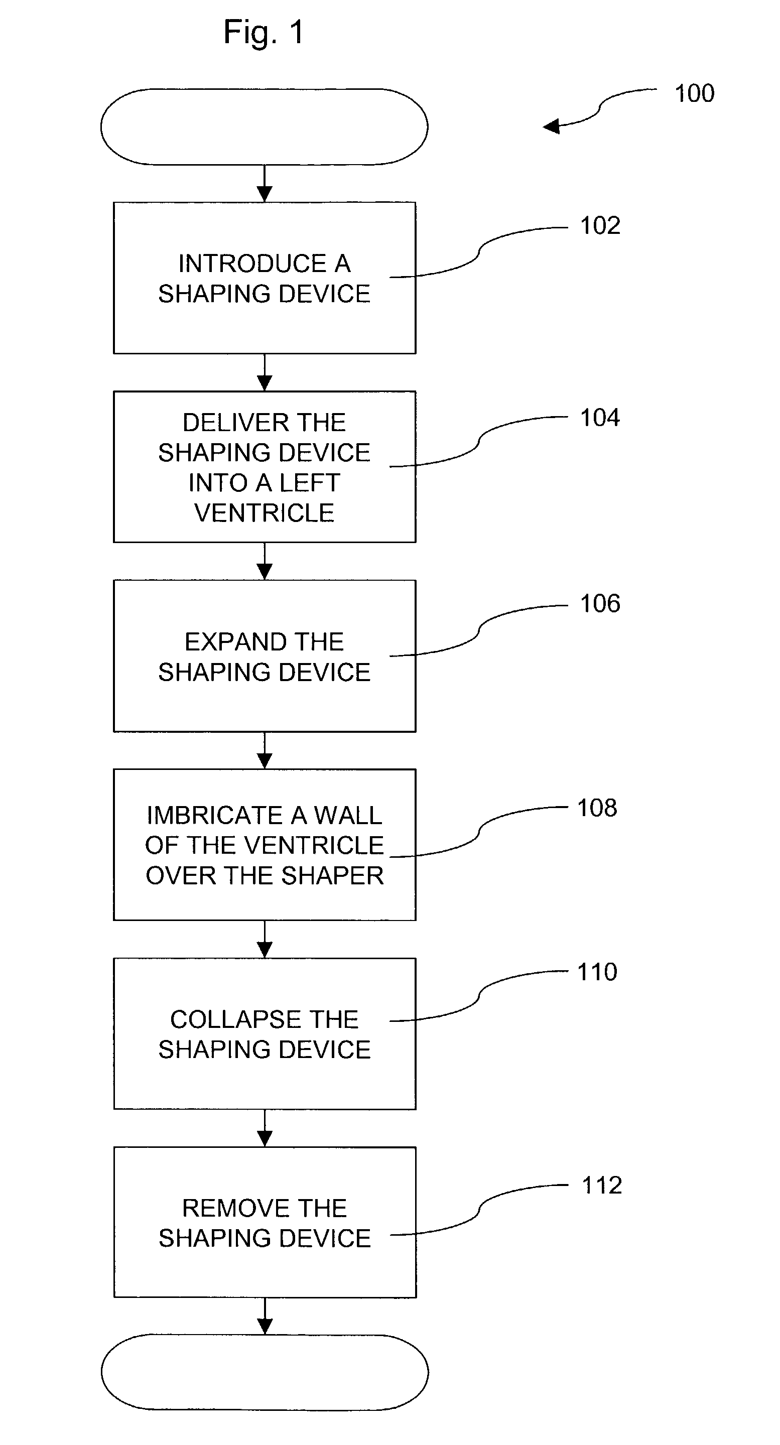
[0034]Turning to FIG. 1, there is presented an overview method 100 for performing and using one embodiment. The method 100 may use the following components: a shaping device, a patch, and a stapling device.

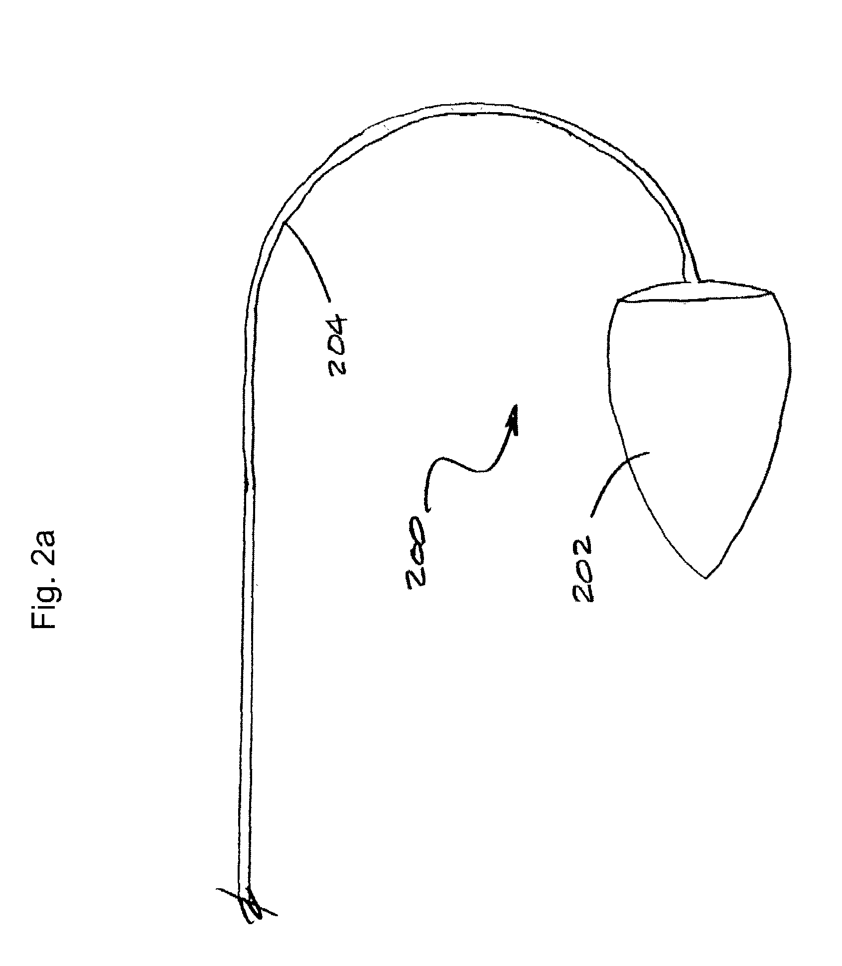
[0035]The shaping device may be pre-shaped to generally model the appropriate volume and shape of the left ventricle, such as illustrated in FIG. 2a. Such a shaping device 200 may be used as a guide in reforming the left ventricle so that the reconstructed heart may be formed closer to the size and shape of the pre-enlarged heart. Consequently, the heart performs better post operatively than with conventional methods. As illustrated in FIG. 2a, the shaping device 200 is generally conical or “tear drop” in shape. The length of the shaping device 200 may vary with each patient and will typically be a function of the volume selected for the shaping device. Depending on the patient, the length may be in the three to four inch range to generally match the length of the pre-enlarged lef...


PUM


Abstract

In one embodiment, there is disclosed a method for repairing a heart of a human, comprising introducing a collapsed shaping device through the skin into the vascular system of the human, delivering the shaping device into a left ventricle through the arteries, once inside the left ventricle, expanding the shaping device to an expanded shape, imbricating a wall of the ventricle over the shaping device, collapsing the shaping device, and removing the shaping device from the left ventricle such that the ventricle is restored to an appropriate size.

Description

PRIORITY CLAIM[0001]This application claims the benefit of the filing date of U.S. provisional patent application Ser. No. 60 / 317,197 entitled “DEVICE AND METHOD FOR ENDOSCOPIC SURGICAL VENTRICULAR REPAIR” filed on Sep. 5, 2001 and U.S. provisional patent 60 / 327,221 entitled “METHOD AND DEVICE FOR CLOSED CHEST PLACEMENT OF SEPTUM” filed on Oct. 5, 2001, the disclosures of both of these applications are incorporated herein by reference.BACKGROUND OF THE INVENTION[0002]1. Field of the Invention[0003]This invention relates generally to surgical methods and apparatuses for performing surgical ventricular repair endoscopically or through a minimally invasive incision.[0004]2. Description of the Related Art[0005]The function of a heart in an animal is primarily to deliver life-supporting oxygenated blood to tissue throughout the body. This function is accomplished in four stages, each relating to a particular chamber of the heart. Initially deoxygenated blood is received in the right auri...

Claims


Application Information

Patent Timeline
IPC IPC(8): A61F2/00A61F13/00
CPCA61F2/2487
Inventor MURPHY, GREGORYSURESH, MITTADAVIS, ALBERT
Owner HEART FAILURE TECH
Why Eureka
Try Eureka