Reconfigurable, fault tolerant multiple-electrode cardiac lead systems

a multi-electrode, fault-tolerant technology, applied in the field of reconfigurable, fault-tolerant medical electrical lead systems, can solve the problems of reduced ventricular asynchrony, hemodynamic insufficiency may require clinical intervention, and reduced ventricular contractile strength ability, etc., to achieve the effect of complex

Inactive Publication Date: 2005-04-28
MEDTRONIC INC
View PDF33 Cites 175 Cited by
  • Summary
  • Abstract
  • Description
  • Claims
  • Application Information

AI Technical Summary

Benefits of technology

[0014] For example, by practicing the methods of the present invention an MI is readily detectable and, as a result, the operative electrodes delivering therapy to or near the region of the MI can be rerouted to effectively deliver therapy elsewhere while avoiding the MI-affected region of the heart.
[0015] In addition, the present invention provides a compact and convenient apparatus for performing a localized study of dispersion of depolarization and repolarization wavefronts within cardiac tissue. The velocity and direction of such wavefronts provides valuable information of the topography of conduction of the myocardium. Such information provides direct evidence of conduction anomalies without requiring visual, echocardiographic or tomographic inspection. That is, a clinician need not perform machine vision analysis (including electrophysiology study, echocardiographic examination, SPECT, NMR, MRI or PET scans, fluoroscopy exposure and the like) and accordingly does not need to effectively and physiologically counter conduction anomalies by tailoring therapy delivery based on the current conduction status of a patient. The information inherently incorporates current physiologic parameters reflecting overall autonomic tone of a patient (e.g., regardless of possibly confounding factors such as various cardiac drugs, diet, physical exertion and the like). As a result, an acute episode of cardiac ischemia and / or a (normal) sinus tachycardia due to physical exertion can be identified and, if necessary, rectified.
[0016] When delivering customized pacing therapy, the present invention provides a platform for performing what is referred to herein as “cascaded pacing” is used to cause intra-chamber synchronization of depolarization. Thus in the event that a depolarization of one or more volumes of myocardium precedes (or lags) adjacent volumes of myocardium, delivery of pacing stimulation improves hemodynamic performance. In a similar fashion, such cascaded pacing is employed to terminate arrhythmias with an intrachamber cascade of anti-tachycardia pacing (ATP). In this form of the invention, by decrementing the -pacing intervals for at least some of the electrodes during successive cardiac cycles a source of undesirable depolarization wavefronts may be effectively countered. For example, if an ectopic focus or an accessory pathway were initiating conflicting depolarization wavefronts, such cascaded ATP can effectively “peel back” such wavefronts until they diminish or are resynchronized with the desired depolarization timing in a chamber.
[0018] Moreover, in the event that one or more of the plurality of electrodes is rendered ineffective or inoperable due to disengagement from adjacent myocardial tissue, electrical open or short circuit condition, or non-optimal disposition the affected electrodes are eliminated from therapy delivery circuitry (and / or cardiac activity sensing circuitry). This aspect of the present invention offers a modicum of fault tolerance so that therapy delivery may continue unimpeded, possibly indefinitely.
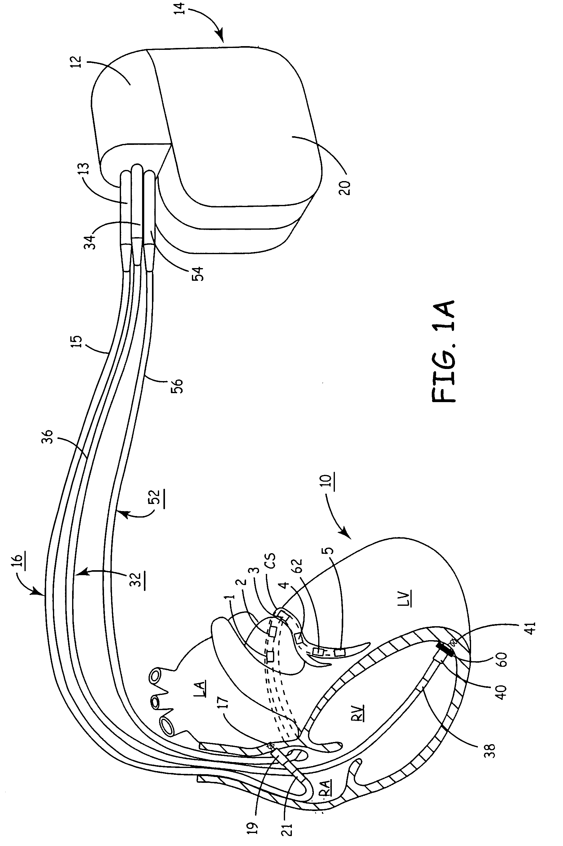
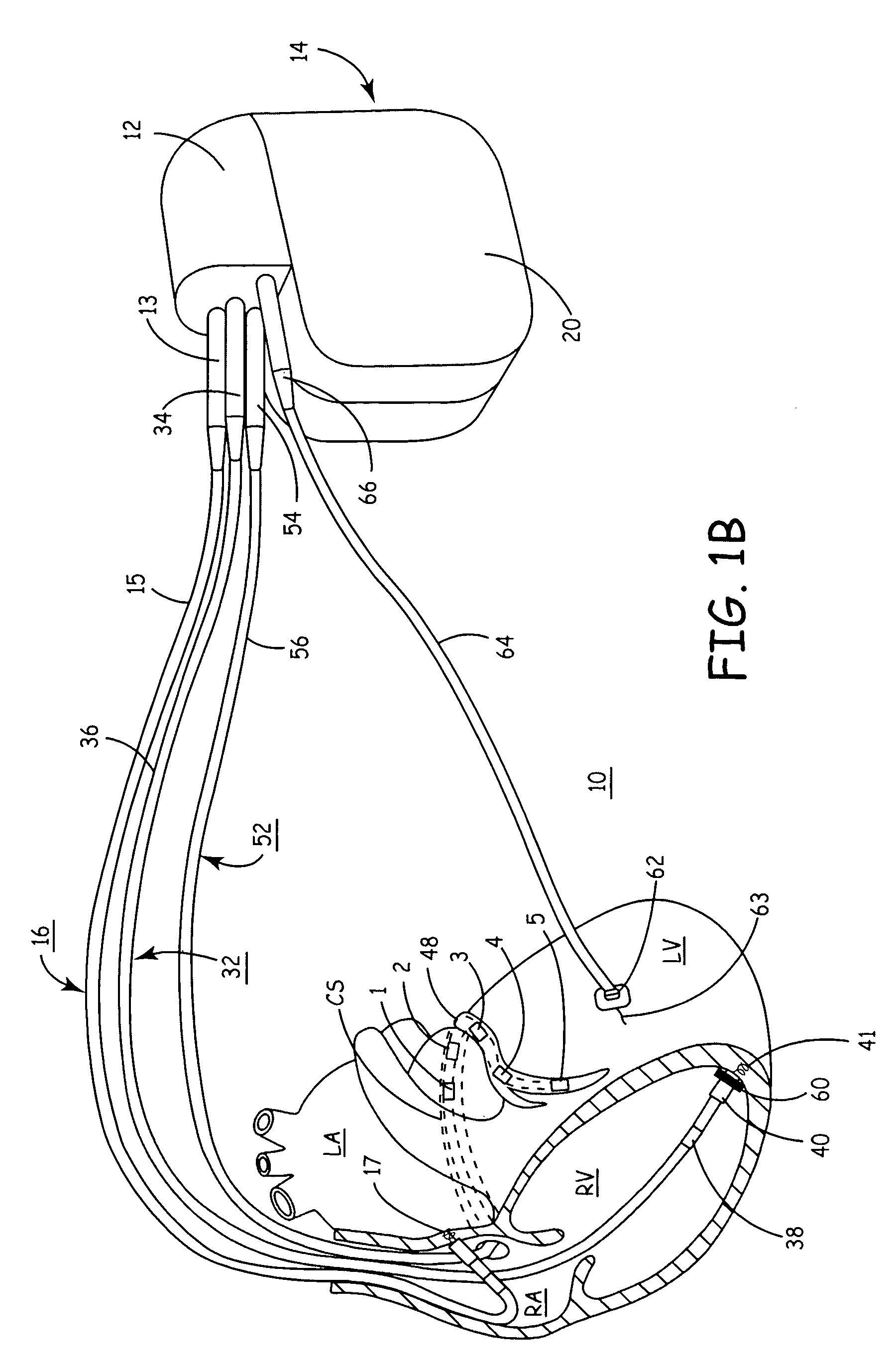
[0023] As is well known, if an output signal from an accelerometer is mathematically integrated the velocity of the adjacent cardiac tissue is measured and displacement signals are available by performing a double mathematical integration of an acceleration signal. Comparing a prior and a recent displacement signal from an accelerometer disposed on the left lateral free wall (preferably disposed about the mid-basal portion of the LV), thereby providing an indirect measurement of LV volume (e.g., comparing systole and diastole volumes). As a result, the present invention provides structure and methods to detect curative effects of CRT and other therapies so that relatively complex (and costly in terms of energy usage) therapeutic regimes may be avoided when not necessary. If the displacement of the LV is reduced significantly, the detected depolarization patterns are relatively uniform, and the LV and RV are contracting in a synchronized manner, CRT delivery may cease with a mode-switch to a more physiologic single- or double-chamber pacing therapy (e.g., AAI, ADI, AAI / R, ADI / R, etc.) without detriment to the patient.

Problems solved by technology

In addition, deleterious remodeling of the myocardium (e.g., LV lateral or “free” wall) oftentimes accompanies HF or other pathological conditions of a patient.
This alteration and / or remodeling, when combined with the effects of weakened cardiac muscles or modified depolarization patterns, reduces the ability of the LV to generate contractile strength.
Given prior art devices and methods, the resulting hemodynamic insufficiency may require clinical intervention.
Ventricular asynchrony following coronary artery bypass graft (CABG) surgery is a problem encountered relatively often, requiring post-operative temporary pacing.
However, not all patients respond favorably to CRT.
Physicians are challenged in selecting patients that will benefit and in selecting the optimal pacing locations and pacing intervals applied to resynchronize the heart chamber contractions.
The foregoing physiologic issues can negatively affect the efficacious delivery of diverse cardiac stimulation therapies.
Significant hemodynamic changes may not always be acutely observable in an individual patient, however, using non-invasive monitoring methods.
As a result, an electrical therapy that previously produced effective results (e.g., adequate cardiac output, stroke volume and cardiac perfusion) can be rendered ineffective.

Method used


Image

  • Reconfigurable, fault tolerant multiple-electrode cardiac lead systems
  • Reconfigurable, fault tolerant multiple-electrode cardiac lead systems
  • Reconfigurable, fault tolerant multiple-electrode cardiac lead systems

Examples


Embodiment Construction

[0044] The present invention provides a method and apparatus for assessing ventricular function on a chronic basis using a plurality of electrodes disposed in operative electrical communication proximate the LV. For certain methods according to the present invention at least one remote electrode disposed outside the LV is desirable (e.g., a canister-based, subcutaneous, pericardial, epicardial, coil type, RA, LA, or RV electrode, etc.). Optionally, at least one mechanical or metabolic sensor also operatively electrically couples to an implantable medical device (IMD) and provides cardiac performance information which can be used in conjunction with the multiple electrode lead system of the present invention.

[0045] The plurality of electrodes are spaced-apart so that a single electrode is disposed in electrical communication with a discrete volume of ventricular tissue. In one embodiment, the discrete volume of tissue is divided by planes as is known and used in the medical arts (e....


PUM


Abstract

The present invention provides a method and apparatus for assessing ventricular function on a chronic basis using a plurality of electrodes disposed on or about a left ventricle and / or a right ventricle—and optionally, at least one mechanical or metabolic sensor—all operatively electrically coupled to an implantable medical device. The plurality of electrodes are preferably spaced-apart so that at least one electrode is disposed electrical communication with a discrete volume of ventricular tissue. In one embodiment, the discrete volume of tissue is defined by multiple longitudinal and axial planes as known and used in the medical arts. Thus, according to the present invention, at least one electrode couples to appropriate sensing circuitry and essentially provides a localized electrogram (EGM) that, when compared to other EGMs, provides for configurable, localized delivery of therapeutic pacing stimulus, diverse impedance-sensing vectors, various diagnostic information regarding myocardial function and / or anti-tachycardia pacing.

Description

CROSS-REFERENCE TO RELATED APPLICATIONS [0001] This patent application relates to and hereby incorporates by reference the contents of non-provisional U.S. patent application Ser. No. 10 / xxx,xxx (Atty. Dkt. P-11118.00) filed on 14 Oct. 2003 and entitled, “METHOD AND APPARATUS FOR MONITORING TISSUE FLUID CONTENT FOR USE IN AN IMPLANTABLE CARDIAC DEVICE,” and non-provisional U.S. patent application Ser. No. 10 / xxx,xxx (Atty. Dkt. P-11193.00) filed 7 Oct. 2003 and entitled, “MULTIPLE PACING OUTPUT CHANNELS.”FIELD OF THE INVENTION [0002] The present invention relates generally to reconfigurable, fault-tolerant medical electrical lead systems for cardiac medical devices. More particularly, the present invention relates to lead systems capable of sensing cardiac events, measuring intrathoracic impedance, and / or delivering diverse electrical stimulation therapies. Optionally, lead systems according to the present invention include mechanical and / or metabolic sensor input to assist in sensi...

Claims


Application Information

Patent Timeline
Patent Type & Authority Applications(United States)
IPC IPC(8): A61B5/363A61N1/05A61N1/365
CPCA61B5/0422A61B5/0464A61N2001/0585A61N1/0587A61N1/36514A61N1/056A61B5/287A61B5/363
Inventor HINE, DOUGLAS S.MANDA, VENSOMMER, JOHN L.
Owner MEDTRONIC INC
Why Eureka
Try Eureka